Chapter 15 Enzyme Regulation
Chapter 15 Enzyme Regulation. Outline. What factors influence enzymatic activity? What are the general features of allosteric regulation? Can allosteric regulation be explained by conformational changes in proteins? What kinds of covalent modification regulate the activity of enzymes?

Chapter 15 Enzyme Regulation
E N D
Presentation Transcript
Outline • What factors influence enzymatic activity? • What are the general features of allosteric regulation? • Can allosteric regulation be explained by conformational changes in proteins? • What kinds of covalent modification regulate the activity of enzymes? • Is the activity of some enzymes controlled by both allosteric regulation and covalent modification? • Special focus: is there an example in nature that exemplifies the relationship between quaternary structure and the emergence of allosteric properties? Hemoglobin and Myoglobin – paradigms of protein structure and function
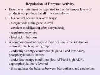
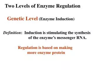
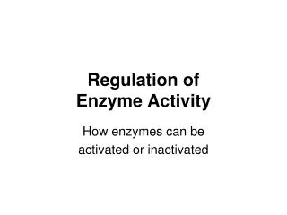
15.1 – What Factors Influence Enzymatic Activity? • The availability of substrates and cofactors usually determines how fast the reaction goes • As product accumulates, the apparent rate of the enzymatic reaction will decrease • Genetic regulation of enzyme synthesis and decay determines the amount of enzyme present at any moment • Enzyme activity can be regulated allosterically • Enzyme activity can be regulated through covalent modification • Zymogens, isozymes, and modulator proteins may play a role
15.1 – What Factors Influence Enzymatic Activity? Enzyme regulation by reversible covalent modification.
15.1 – What Factors Influence Enzymatic Activity? Zymogens are inactive precursors of enzymes. Typically, proteolytic cleavage produces the active enzyme. Proinsulin is an 86-residue precursor to insulin
Isozymes Are Enzymes With Slightly Different Subunits The isozymes of lactate dehydrogenase (LDH).
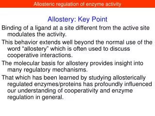
15.2 – What Are the General Features of Allosteric Regulation? Action at "another site" • Enzymes situated at key steps in metabolic pathways are modulated by allosteric effectors • These effectors are usually produced elsewhere in the pathway • Effectors may be feed-forward activators or feedback inhibitors • Kinetics are sigmoid ("S-shaped")
15.2 – What Are the General Features of Allosteric Regulation? Sigmoid v versus [S] plot. The dotted line represents the hyperbolic plot characteristic of normal Michaelis=Menten kinetics.
15.3 Can Allosteric Regulation Be Explained by Conformational Changes in Proteins? Monod, Wyman, Changeux (MWC) Model: - Allosteric proteins can exist in two states: R (relaxed) and T (taut) - In this model, all the subunits of an oligomer must be in the same state - T state predominates in the absence of substrate S - S binds much tighter to R than to T
The Symmetry Model for Allosteric Regulation is Based on Two Conformational States for a Protein Allosteric effects: A and I binding to R and T, respectively.
The Symmetry Model for Allosteric Regulation is Based on Two Conformational States for a Protein Allosteric effects: A and I binding to R and T, respectively.
The Symmetry Model for Allosteric Regulation is Based on Two Conformational States for a Protein
More about the MWC model • Cooperativity is achieved because S binding increases the population of R, which increases the sites available to S • Ligands such as S are positive homotropic effectors • Molecules that influence the binding of something other than themselves are heterotropic effectors
The Sequential Model for Allosteric Regulation is Based on Ligand-Induced Conformation Changes • An alternative model – proposed by Koshland, Nemethy, and Filmer (the KNF model) relies on the idea that ligand binding triggers a conformation change in a protein • If the protein is oligomeric, ligand-induced conformation changes in one subunit may lead to conformation changes in adjacent subunits • The KNF model explains how ligand-induced conformation changes could cause subunits to adopt conformations with little affinity for the ligand – i.e., negative cooperativity • The KNF model is termed the sequential model
The Sequential Model for Allosteric Regulation is Based on Ligand-Induced Conformation Changes The Koshland-Nemethy-Filmer sequential model for allosteric behavior. (a) S binding can, by induced fit, cause a conformational change in the subunit to which it binds. (b) If subunit interactions are tightly coupled, binding of S to one subunit may cause the other subunit to assume a conformation having a greater or lesser affinity for S. That is, the ligand-induced conformational change in one subunit can affect the adjoining subunit.
The Sequential Model for Allosteric Regulation is Based on Ligand-Induced Conformation Changes The Koshland-Nemethy-Filmer model. Theoretical curves for the binding of a ligand to a protein having four identical subunits, each with one binding site for the ligand.
The notable difference between MWC and KNF models • In the MWC model, the different conformations have different affinities for the various ligands, and the concept of ligand-induced conformational changes is ignored • In contrast, the KNF model is based on ligand-induced conformational changes
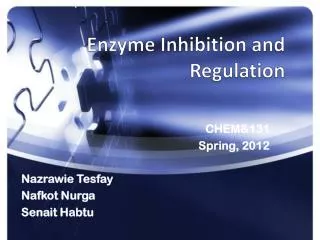
15.4 What Kinds of Covalent Modification Regulate the Activity of Enzymes? • Enzyme activity can be regulated through reversible phosphorylation • This is the most prominent form of covalent modification in cellular regulation • Phosphorylation is accomplished by protein kinases • Each protein kinase targets specific proteins for phosphorylation • Phosphoprotein phosphatases catalyze the reverse reaction – removing phosphoryl groups from proteins • Kinases and phosphatases themselves are targets of regulation
15.4 What Kinds of Covalent Modification Regulate the Activity of Enzymes? • Protein kinases phosphorylate Ser, Thr, and Tyr residues in target proteins • Kinases typically recognize specific amino acid sequences in their targets • In spite of this specificity, all kinases share a common catalytic mechanism based on a conserved core kinase domain of about 260 residues (see Figure 15.9) • Kinases are often regulated by intrasteric control, in which a regulatory subunit (or domain) has a pseudosubstrate sequence that mimics the target sequence, minus the phosphorylatable residue
15.4 What Kinds of Covalent Modification Regulate the Activity of Enzymes?
Cyclic AMP-dependent protein kinase is composed of catalytic and regulatory subunits cyclic AMP-dependent protein kinase (also known as protein kinase A (PKA) is a 150- to 170-kD R2C2 tetramer in mammalian cells. The two R (regulatory) subunits bind cAMP; cAMP binding releases the R subunits from the C (catalytic) subunits. C subunits are enzymatically active as monomers.
Phosphorylation is Not the Only Form of Covalent Modification that Regulates Protein Function • Several hundred different chemical modifications of proteins have been discovered • Only a few of these are used to achieve metabolic regulation through reversible conversion of an enzyme between active and inactive forms • A few are summarized in Table 15.3 • Three of the modifications in Table 15.3 require nucleoside triphosphates (ATP, UTP) that are related to cellular energy status
Phosphorylation is Not the Only Form of Covalent Modification that Regulates Protein Function
Acetylation in Enzyme Regulation • Acetylation is a prominent modification for the regulation of metabolic enzymes • Acetylation of an ε-NH3+ group on a Lys residue changes it from a positively charged amino group to a neutral amide • This change may have consequences for protein structure and thus function • The acetylating enzyme is termed an acetyl-CoA-dependent lysine acetyltransferase or KAT • More than 30 KATs are known in mammals • Deacetylation by KDACs (lysine deacetylases) reverse the effects of acetylation
Acetylation in Enzyme Regulation • Proteomics studies show that acetylation of metabolic enzymes is an important mechanism for regulating the flow of metabolic substrates (carbohydrates and fats, for example) through the central metabolic pathways • Acetylation activates some enzymes and inhibits others • Cellular levels of major metabolic fuels such as glucose, fatty acids, and amino acids influence the degree of acetylation • The KDACs include sirtuins, a class of NAD+-dependent protein deacetylating enzymes • Sirtuins are implicated in energy metabolism and longevity
15.5 Are Some Enzymes Controlled by Both Allosteric Regulation and Covalent Modification? • Glycogen phosphorylase (GP) is an example of the many enzymes that are regulated both by allosteric controls and by covalent modification • GP cleaves glucose units from nonreducing ends of glycogen • This converts glycogen into readily usable fuel in the form of glucose-1-phosphate • This is a phosphorolysis reaction • Muscle GP is a dimer of identical subunits, each with PLP covalently linked • There is an allosteric effector site at the subunit interface
Glycogen Phosphorylase is Controlled by Both Allosteric Regulation and Covalent Modification The mechanism of covalent modification and allosteric regulation of glycogen phosphorylase.
Glycogen phosphoryase is activated by a cascade of reactions The hormone-activated enzymatic cascade that leads to activation of glycogen phosphorylase.
Hemoglobin A classic example of allostery • Hemoglobin and myoglobin are oxygen- transport and oxygen-storage proteins, respectively • Compare the oxygen-binding curves for hemoglobin and myoglobin • Myoglobin is monomeric; hemoglobin is tetrameric • Mb: 153 aa, 17,200 MW • Hb: two α chains of 141 residues, 2 β chains of 146 residues
The structure of myoglobin is similar to that of an Hb monomer The myoglobin and hemoglobin structures. Myoglobin is monomeric Hemoglobin is tetrameric
Mb and Hb use heme to bind Fe2+ Heme is formed when protoporphyrin IX binds Fe2+
Fe2+ is coordinated by His F8 • Iron interacts with six ligands in Hb and Mb • Four of these are the N atoms of the porphyrin • A fifth ligand is donated by the imidazole side chain of amino acid residue His F8 • (This residue is on the sixth or “F” helix, and it is the 8th residue in the helix, thus the name.) • When Mb or Hb bind oxygen, the O2 molecule adds to the heme iron as the sixth ligand • The O2 molecule is tilted relative to a perpendicular to the heme plane
Fe2+ is coordinated by His F8 The six liganding positions of an iron atom in Hb and Mb.
Myoglobin Structure • Mb is a monomeric heme protein • Mb polypeptide "cradles" the heme group • Fe in Mb is Fe2+ - ferrous iron - the form that binds oxygen • Oxidation of Fe yields 3+ charge - ferric iron • Mb with Fe3+ is called metmyoglobin and does not bind oxygen
O2 Binding Alters Mb Conformation • In deoxymyoglobin, the ferrous ion actually lies 0.055 nm above the plane of the heme • When oxygen binds to Fe in heme of Mb, the heme Fe is drawn toward the plane of the porphyrin ring • With oxygen bound, the Fe2+ atom is only 0.026 nm above the plane • For Mb, this small change has little consequence • But a similar change in Hb initiates a series of conformational changes that are transmitted to adjacent subunits
Hb Has an α2β2 Tetrameric Structure An αβ dimer of Hb, with packing contacts indicated in blue. The sliding contacts made with the other dimer are shown in yellow.
Cooperative Binding of Oxygen Influences Hemoglobin Function • Mb, an oxygen-storage protein, has a greater affinity for oxygen at all oxygen pressures • Hb is different – it must bind oxygen in lungs and release it in capillaries • Hb becomes saturated with O2 in the lungs, where the partial pressure of O2 is about 100 torr • In capillaries, pO2 is about 40 torr, and oxygen is released from Hb • The binding of O2 to Hb is cooperative – binding of oxygen to the first subunit makes binding to the other subunits more favorable
O2-Binding Curves of Mb and Hb The oxygen binding curve of Mb resembles an enzyme:substrate saturation curve.
An Alternative O2-Binding Curve for Hb Oxygen saturation curve for Hb in the form of Y versus pO2 assuming n=4 and P50 =26 torr. Y is the fractional saturation of Hb:
An Alternative O2-Binding Curve for Hb A comparison of the experimentally observed O2 curve for Hb yielding a value for n of 2.8, the hypothetical curve if n=4, and the curve if n=1 (non-interacting O2-binding sites).
The Conformation Change The secret of Mb and Hb • Oxygen binding changes the Mb conformation • Without oxygen bound, Fe2+ is out of heme plane • Oxygen binding pulls the Fe2+ into the heme plane • Fe2+ pulls its His F8 ligand along with it • The F helix moves when oxygen binds • Total movement of Fe2+ is 0.029 nm – i.e., 0.29 Å • This change means little to Mb, but lots to Hb!
Oxygen Binding by Hb Induces a Quaternary Structure Change • When deoxy-Hb crystals are exposed to oxygen, they shatter. Evidence of a large-scale structural change • One alpha-beta pair moves relative to the other by 15 degrees upon oxygen binding • This massive change is induced by movement of Fe by 0.039 nm when oxygen binds
Oxygen binding to Hb results in a 15° rotation of one αβ pair relative to the other Subunit motion in hemoglobin when the molecule goes from the (a) deoxy form to the (b) oxy form.
Fe2+ Movement by Less Than 0.04 nm Induces the Conformation Change in Hb • In deoxy-Hb, the iron atom lies out of the heme plane by about 0.06 nm • Upon O2 binding, the Fe2+ atom moves about 0.039 nm closer to the plane of the heme • It is as if the O2 is drawing the heme iron into the plane • This may seem like a trivial change, but its biological consequences are far-reaching • As Fe2+ moves, it drags His F8 and the F helix with it • This change is transmitted to the subunit interfaces, where conformation changes lead to the rupture of salt bridges
Fe2+ Movement by Less Than 0.04 nm Induces the Conformation Change in Hb Changes in the position of the heme iron atom upon oxygenation lead to conformational changes in the hemoglobin molecule.
The Physiological Significance of the Hb:O2 Interaction • Hb must be able to bind oxygen in the lungs • Hb must be able to release oxygen in capillaries • If Hb behaved like Mb, very little oxygen would be released in capillaries • The sigmoid, cooperative oxygen-binding curve of Hb makes its physiological actions possible!
H+ Promotes Dissociation of Oxygen from Hemoglobin • Binding of O2 to Hb is affected by several agents, including H+, CO2, and chloride ions • The effect of H+ is particularly important • Deoxy-Hb has a higher affinity for H+ than oxy-Hb • Thus, as pH decreases, dissociation of O2 from hemoglobin is enhanced • Ignoring the stoichiometry of O2 and H+, we can write: HbO2 + H+ HbH+ + CO2
H+ Promotes Dissociation of Oxygen from Hemoglobin The oxygen saturation curves for myoglobin and for hemoglobin at five different pH values: 7.6, 7.4,7.2, 7.0, 6.8.