Download
1 / 64
650 likes | 685 Views
Dive into the fascinating world of cells with this comprehensive guide covering cell theory, the history of microscopy, different types of cells, and their structures and functions. Learn about pioneers like Anton Van Leeuwenhoek and Robert Hooke, discover the principles of complementarity of structure and function, and explore the main cell structures and organelles. Unveil the Fluid Mosaic Model of cell membranes and understand the role of plasma membranes, membrane proteins, and cytoplasm in cellular function.

E N D
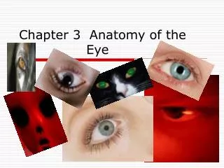
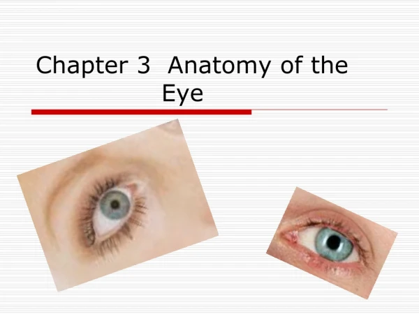
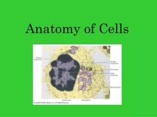
Chapter 3 Anatomy of Cells
Cell Theory • All living things are composed of one or more cells. • Cells are the basic units of structure and function in living things. • New cells are produced from existing cells.
In 1839 German Scientists Theodor Schwann and Matthias Schleiden suggested that cells were the basic unit of life and that all living things came from cells
Functional Anatomy • Refers to study of structure as they relate to function
Light Microscope • Also referred to as the optical microscope • Uses visible light and a system of lenses to magnify images of small samples. • Limit of 2000 times greater Skin cells
Anton Van Leeuwenhoek • Dutchman • Commonly known as "the Father of Microbiology“ • Considered to be the first microbiologist. • Didn’t invent but did improve the light microscope. • He was first to observed single celled organisms which he called animalcules
Robert Hooke • Englishman • Known as "the father of microscopy“ • Coined the term "cell" due to the fact that what he was looking at reminded him of a monk’s cell in a monastery • Published a book in 1665 titled Micrographia
Electron Microscope • Uses a particle beam of electrons to illuminate a specimen and create a highly-magnified image. • Can obtain much higher magnifications of up to 2 million times. • First prototype built in 1931 Fly foot
Transmission Electron MicroscopeTEM • Electrons are emitted by an electron gun • Images are called transmission electron micrographs Hair
Scanning Electron MicroscopeSEM • Beam of electrons scan the surface and are reflected from the surface • Gives illusion of depth Single hair cell from frog’s ear Dust mite
Principle of Complementarity of Structure and Function • An organ's structure cannot be studied without understanding how it functions in the living system • An organ's function cannot be studied without understanding it’s structure in the living system.
Nerve Cell • Surface sensitive to stimuli • Long extensions • Detects changes in internal or external environment • Transmit nerve impulses from one part of the body to another
Muscle Cell • Elongated and threadlike • Has tiny fibers that slide together forcefully • Made to contract or shorten • Used in movement of body parts
Red Blood Cells • Contains hemoglobin • Transports oxygen in the blood stream RBC with malaria
Gland Cells • Contain sacs that release secretion to outside of the cells • Releases substances such as hormones, enzymes, mucous and sweat. Exocrine gland
Immune Cells • Main function is to destroy “nonself” cells such as cancer or bacteria. • Some have outer membranes that can engulf other cells • Some can manufacture antibodies • Some can destroy other cells T cell
Composite Cell • Typical cell • Cells have many similarities
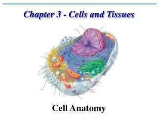
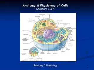
3 Main Cell Structures • Plasma membrane • Cytoplasm • Nucleus
Cell Structures • Plasma membrane surrounds the cell • Inside cell is composed gel-like substance called cytoplasm • Cytoplasm is made up of various organelles and a watery fluid called cytosol or intracellular fluid • Nucleus is in the center of cell and is not part of cytoplasm.
Review • What important concept was purposed by Schleiden and Schwann? • Give example of how cell structure relates to its function. • Three main cell structures
Cell Membranes and Organelles • Made of lipids, protein and other molecules • Each cell has various membranous organelles which are sacs and canals made from the same material as the plasma membrane (See Table 3-2 page 77)
Membranous Structures • Plasma membrane • Endoplasmic Reticulum (ER) • Golgi Apparatus • Lysosomes • Peroxisomes • Mitochondria • Nucleus
Nonmembranous Structures • Ribosomes • Cytoskeleton • Cilia and flagella • Nucleolus
Fluid Mosaic Model • Model of cell membrane structure composed of a lipid bilayer with scattered proteins; often described as a sea of lipids with protein icebergs. Fluid and moves around with a flowing changing pattern of arrangement.
Plasma Membrane/Cell Membrane • Bi-layer of phospholipids • Hydrophobic tails and Hydrophilic heads
Cholesterol • Steroid lipid that mixes with the phospholipids. • Stays fluid enough to function at body temperature. • Without it cell membranes would break far too easily • Forms fence like material that allows lipid soluble molecules to pass through
Membrane Proteins • Act like gates allowing water soluble molecules through membranes. • Glycoproteins are formed • Glycoproteins have carbohydrates attached to them acting like identification markers • Some are receptors that react to things such as hormones • Carry molecules across membrane in a process known as signal transduction • See Table 3-3 page 80
Cytoplasm • Gel-like internal substance • Contains suspended structures • Highly differentiated into organelles and cytosol
Endoplasmic Reticulum (ER) • Endoplasm means cytoplasm is located toward the center of the cell • Reticulum means network • ER however is located throughout the cell as seen by electron microscopes • ER walls are basically the same molecular structure as the plasma membrane • Function in protein synthesis
Endoplasmic Reticulum (ER) • Allows intracellular transport of molecules • Proteins move through canals toward the golgi apparatus • Two types: Rough and Smooth
Rough ER • Ribosomes on the surface of the membrane make it rough • Proteins are synthesized, enter the canal and move toward the Golgi apparatus and eventually leave cell.
Smooth ER • No ribosomes on walls • Smooth ER believed to synthesize lipids and carbohydrates
Ribosomes • Protein factories: Protein synthesis • Many are attached to rough ER • Can not be seen with a light microscope • Made of two subunits. A large one and a small one (figure 3-6 page 82)
Ribosomes • Each subunit contains a ribonucleic acid (RNA) bonded to a protein (rRNA, mRNA, tRNA) • Working ribosomes function in groups called polyribosomes. • Polyribosomes look like a string of beads under the electron microscope.
Golgi Apparatus • It is a membranous organelle • Has several canals called cisternae • Process and packages protein molecules for export out of cell • Proteins are packaged into membranous bubbles called vesicles
Lysosomes • Lysosomes have membranous walls. • Vesicles that have been pinched off from the Golgi apparatus • Filled with enzymes capable of breaking down cell components • Lysosomes destroy cells by digesting them • They are the cellular garbage disposal of the cell • White blood cells
Peroxisomes • Membranous organelle • Detoxify the cell • Contain peroxidase and catalase
Mitochondria • Membranous organelle • Called “the power house” of the cell • Produce ATP for cell energy use • Have 2 membrane walls • Inner wall folded into cisternae • Outer membrane has enzymes in it • Number of mitochondria in a cell has been linked to the cell’s activity
Other Organelles • Box 3-2 Page 81 • Originally just listed as inclusions • Barrel • Compartment for Peptide Loading (CPL) • Caveola
Review • List 3 functions of the plasma membrane • Identify 3 organelle and give functions • Distinguish between membranous and nonmembranous structures
Cytoskeleton • Cell’s internal supporting framework • Made up of rod-like pieces that support and allow movement • Has muscle-like groups of fibers that move the cell or cell parts
Cell Fibers • Form 3D irregularly shaped lattice or scaffolding • Appear to support ER, mitochondria and free floating ribosomes • They have microfilaments, intermediate filaments and microtubules • See page 85
Microfilaments • Smallest cell fibers • Often serve as “cellular muscle” • Made of thin twisted strands of protein molecules • Some can slide past one another causing shortening of the cell, such as in muscle fibers