Download
1 / 32
320 likes | 356 Views
Learn about the anatomy of cells, including cellular components, structure, and function. Explore the typical cell, plasma membrane, cytoplasm, organelles, endoplasmic reticulum, ribosomes, Golgi apparatus, lysosomes, and mitochondria. Understand the role of the nucleus, nuclear structure, and the cytoskeleton in cell biology. Enhance your knowledge of cell biology with this comprehensive guide.

E N D
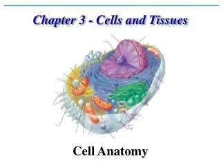
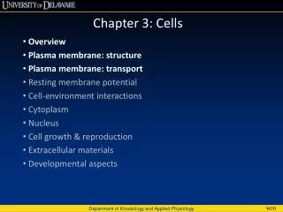
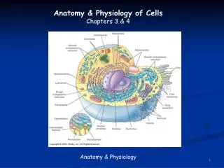
Anatomy of Cells • Cell Structure • Cellular Components • Structure • Function
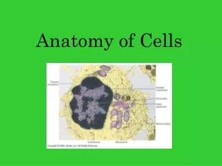
The typical cell (Figure 3-1) – generic cell Varies in size; all are microscopic (Table 3-1) Varies in structure and function (Table 3-2) Functional Anatomy of Cells
Cell Structures • Plasma membrane—separates the cell from its surrounding environment • Cytoplasm—thick gel-like substance inside of the cell composed of numerous organelles suspended in watery cytosol; each type of organelle (“little organ”) is suited to perform particular functions (Figure 3-2) • Nucleus—large membranous structure near the center of the cell
Cell Membranes • Plasma membrane (Figure 3-3) • Membranous organelles – sacs and canals made of the same material as the plasma membrane
Cell Membranes • Structure – is a double layer of phospholipid molecule • Phospholipid • Heads are hydrophilic (water-loving) • Tails are hydrophobic (water-fearing) • Cholesterol molecules are scattered among the phospholipids to allow the membrane to function properly at body temperature • Membrane Proteins • Controls what moves through the membrane • Act as i.d. markers • Act as receptors
Cell Membrane • Membrane Function • To keep cellular components inside the cell and extracellular material outside the cell • Controls what moves into and out of the cell
Cytoplasm and Organelles • Cytoplasm – gel-like internal substance of cells that includes many organelles suspended in watery intracellular fluid called cytosol • Cytosol – the watery intracellular fluid • Organelles – “little organs” each have a particular structure and function • Know the function of each organelle and be able to identify it in a generalized figure of the cell
Endoplasmic Reticulum (Figure 3-5) • Function: Synthesis of proteins that will be excreted from the cell (rough ER) and synthesize lipids for the cell membrane, steroid hormones, and certain carbohydrates, removes and stores Ca2+ from the cell’s interior
Endoplasmic Reticulum • Two types of ER: • Smooth ER – do not have ribosomes attached • Synthesizes certain lipids and carbohydrates and creates membranes for use throughout cell • Removes and stores Ca++ from cell’s interior. • Rough ER – have ribosomes attached to the outer surface • Ribosomes synthesize proteins, which move toward the Golgi apparatus and then eventually leave the cell • Function in protein synthesis and intracellular transportation
Ribosomes • Function: the site of protein synthesis • Attached to rough ER or scattered in the cytoplasm • Structure: made of two pieces, a large subunit and a small subunit • Ribosomes in the endoplasmic reticulum make proteins for “export” or to be embedded in the plasma membrane; free ribosomes make proteins for the cell’s domestic use
Golgi Apparatus • Function: Synthesizes carbohydrates, processes proteins from the ER; the cell’s “post office” • Structure: cisternae stacked on one another and located near the nucleus • Processes protein molecules from the endoplasmic reticulum (Figure 3-8)
Lysosomes • Function: Bags of digestive enzymes break down defective cell parts and ingested particles; a cell’s “digestive system” • Structure: Made of microscopic membranous sacs that have “pinched off” from Golgi apparatus
Proteasomes • Function: Hollow protein cylinders that break down abnormal/misfolded proteins and normal proteins no longer needed by the cell
Peroxisomes • Function: contain enzymes that detoxify harmful substances • Often seen in kidney and liver cells
Mitochondria • Function: A cell’s “power plant”; the site of ATP synthesis • Mitochondrial DNA: Each mitochondrion has a DNA molecule, allowing it to produce its own enzymes and replicate copies of itself
Nucleus • Definition—spherical body in center of cell; enclosed by an envelope with many pores • Function: Contains DNA (genetic code) – the “brain” of the cell, dictates protein synthesis
Nuclear Structure • Nuclear Envelope – nuclear membrane, has nuclear pores (controls entrance in and out of the cell) • Nucleoplasm – nuclear substance • Chromatin – the DNA in non-dividing cells • Nucleolous – found in the nucleus, synthesizes rRNA and combines it with protein to form ribosomes
Nucleus • Contains DNA (heredity molecules), which appear as the following: • Chromatin threads or granules in nondividing cells • Chromosomes in early stages of cell division • Functions of nucleus are functions of DNA molecules; DNA determines both structure and function of cells and heredity
Cytoskeleton • Function: acts as a framework to support the cell and its organelles; involved in cell movement; forms cell extensions
Cytoskeleton • Cell fibers – 3 types • Microfilaments • Intermediate Filaments • Microtubules
Microfilaments • Smallest cell fibers • “Cellular muscles” • Made of thin, twisted strands of protein molecules that lie parallel to the long axis of the cell • Microfilaments can slide past each other, causing shortening of the cell
Intermediate Filaments • Twisted protein strands slightly thicker than microfilaments; form much of the supporting framework in many types of cells
Microtubules • Tiny, hollow tubes that are the thickest of the cell fibers • Function: move things around in the cell
Centrosome • Also called the microtubule-organizing center (MTOC) • Plays an important role during cell division • The general location of the centrosome is identified by the centrioles
Cell Extensions • Cytoskeleton that forms projections that extend the plasma membrane outward to form tiny, fingerlike processes
Three Types of Cell Extensions • Microvilli – founds in epithelial cells that line intestines, increase surface area for absoption • Cilia – short and numerous, move substances along the surface of a cell • Flagella – involved in total cell movement; found on human sperm cells
The Big Picture • Review • Conclusions