Download
1 / 38
450 likes | 988 Views
Instrumentation and Detection. Harry M. Johnson, PhD CancerCare Winnipeg, Manitoba, Canada. Instrumentation and Detection. Purpose: To discuss the detection of radiation from the point of view of the classical instrumentation. Instrumentation and Detection: Outline.

E N D
Instrumentation and Detection Harry M. Johnson, PhD CancerCare Winnipeg, Manitoba, Canada
Instrumentation and Detection • Purpose: To discuss the detection of radiation from the point of view of the classical instrumentation.
Instrumentation and Detection: Outline • The Basic Gas-Filled Detector • Classes of Gas-Filled Detectors • The Geiger Tube as an Exposure Meter • Choosing a Radiation meter • Scintillation Detectors • Calibrating a Contamination Meter • Neutron Meter • Thermoluminescent Personnel Dosimetry
The Gas-Filled Detector - 1 • Gas-filled counting chamber • Use air as the gas for initial testing • sealed or unsealed • coaxial electrodes, well insulated • variable voltage, defined anode and cathode • high resistance resistor • capacitance C • radiation enters chamber and ionizes the gas • ions drift towards the electrodes • Voltage pulse is detected
The Gas-Filled Detector - 2 • Apply low voltage • a few ion pairs drift to electrodes • time constant RC is long • Voltage V= Q/C is detected at output • Pulse curve has elongated shape • Difficult to detect successive pulses
The Gas-Filled Detector - 3 • Maintain the same voltage • “drifting” of ion pairs drift to electrodes is the mechanism of charge transport from ion pair production • shorten the time constant RC • Voltage V= Q/C continues to be detected at output • Pulse curve has “clipped” shape • Successive pulses are detectable • Now possible to count the individual pulse • This is the preferred chamber design • Possible to calculate maximum pulse rate
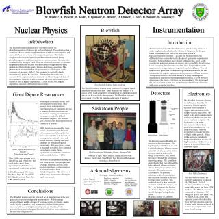
Incident Ionizing Radiation Gas-Filled Detectors Voltage Source + - + + + + Electrical Current Measuring Device - - - - Anode + Cathode - Air or Other Fill Gas
Ion Chamber Instrument • Example of Ion Chamber • 6 cc Chamber • 180 cc Chamber • Readout unit is located remote from the detector
The Gas-Filled Detector • The first plateau region is the “ion chamber” mode • Typically 300 volts applied voltage • Pulse size is independent of LET • No secondary ionizations - which would amplify the pulse height • Pulse size distinction is a disadvantage
The Ionization Chamber • Radiation of constant flux is applied to chamber • Vary the voltage in the few-hundreds of volts region • ionizations in the air of the chamber create ion pairs • a plateau region is noted - the operating plateau for the ion chamber • V is just high enough to collect all ions from ionizations • Ions are not being accelerated - still “drifting” to electrodes • Pulses are distinguishable - each pulse is a single ionization eventPules size is independent of voltage • a beta particle produces 1000 ion pairs, output pulse voltage is low millivolts • Low output voltage is disadvantageous
The Proportional Counter • Adjust the voltage upward beyond the ion chamber plateau • Ions from the initial ion pairs are accelerated • electron acceleration is more important than positive ions • Secondary electrons are produced by collisions by the primary ion-pair products • Change the gas in the chamber • These new design features create a gas amplification factor (>1) - called an “avalanche” • By increasing the tube voltage the avalanche spreads along the anode wire • Pulse size is proportional to the chamber voltage • Pulse size depends on the electric field gradient • Anode (central) diameter is important
The Proportional Counter • This is the “Proportional Counter” Region • Note slight upward slope of Voltage-Pulse curve in this region • Pulse size is dependent on LET - giving the ability to discriminate between type of radiation • Anode wire size is typically 0.02 to 0.1 mm diameter • Decreasing the gas pressure increases the multiplication, although the tube may be at atmospheric pressure • High-voltage supply must be very stable
The Geiger Tube - 1 • Basic Readout module • Two Geiger tubes: “Pancake” type and the “End-Window” type. • Entrance windows are very thin
The Geiger Tube - 2 • Continue to raise the voltage past the “proportional counter” plateau to a new plateau. • Slight upward slope of Voltage-Pulse curve in this region. • Counting rate is independent of voltage. • Change the gas to Argon ( = 15.7 eV; W = 26.4 eV) or Methane ( = 15.2 eV; W = 27.0 eV). • Add 10% ethyl alcohol to eliminate uv . • Tube is sealed. • Chamber pressure may be 10% of atmospheric pressure • Output pulse (without amplification) height:approx 1 volt • Little need for amplification • Output pulses occur independent of size of initiating ionization - no discrimination re LET of the radiation.
The Geiger Tube - 3 • This is Geiger (Geiger-Mueller) tube region (1908). • Voltage is sufficiently high that both ions of the initiating ion pair are accelerated. • Accelerated ions cause additional ionizations (avalanche). • Accelerate +ve ions strike cathode (tube shell), cause excitations of cathode molecules, yielding UV production. • UV is additional source of gas ionization/excitation. • When intense ionization occurs in the tube the E-Field along the anode wire drops to zero. This causes dead time. • GM tube can go into continuous discharge when this occurs. Add alcohol to argon gas to quench the discharge. • Alcohol reduces dead time to 100 micro seconds. (Resolve up to 10,000 pulses/sec). • GM tube is easy to build, simple electronics, cheap.
The Geiger Tube - 4 • Choose operating voltage for G-M region at 1/3 to 1/2 way up the plateau. • Alcohol is present to quench the ionizations and absorb the UV produced when accelerated +ve ions strike shell. • Alcohol molecules dissociate in this process. • Lifetime of the tube is limited by the alcohol - total lifetime is 108 - 109 ionizations. • Ifd voltage is raised above the Geiger region, the avalanche spreads and continuous discharge occurs. Gas tube cannot operate as a detector above the Geiger region.
Resolving Time and Dead Time • Two ionizations in G-M tube in rapid succession may not be resolved. • The first ionization causes a Dead Time when no new pulse can be detected • Followed by Recovery Time when a new pulse may not be identifiable • Resolving Time is sum of Dead and Recovery times
Resolving Time and Dead Time - 2 • Avalanche starts near the anode wire, spreads along anode due to the High Voltage in G-M region • Electrons move more quickly than +ve ions • Rate-limiting step is the transit time of +ve ions to the cathode. This transit time defines the collection time • Resolving time can be defined • Tube resets itself after recovery • Collection time may be a few hundred microseconds • If CT is 250 s, what is limiting rate of detectable ionizations - how many photos per sec is max input?
Advantages/Disadvantages of Gas Detection Tubes • Ion Chamber: simple, accurate, wide range, sensitivity is function of chamber size, no dead time • Proportional Counter: discriminate hi/lo LET, higher sensitivity than ion chamber • GM Tube: cheap, little/no amplification, thin window for low energy; limited life
Geiger Tube as Exposure Meter • “Exposure” is the parameter measuring the ionization of air. • Geiger tube measures ionization pulses per second - a “count rate”. • The number of ionizations in the Geiger tube is a constant for a particular energy but is energy dependent.
Energy Compensation of Geiger Tube • An energy compensating shield is required to smooth out the energy response
Scintillation Detector • Readout Module plus Detector • Photomultiplier Tube (P) • Scintillator Chrystal (C) C P
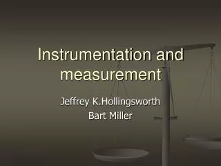
Incident Ionizing Radiation Scintillation Detectors Photomultiplier Tube Pulse Measuring Device Light Photon - Sodium-Iodide Crystal Dynode Anode Photocathode Optical Window
Scintillation Detectors • Construction of Crystal and PMT assembly • Design of basic electronics • Principle of scintillation • Principle of operation
Scintillation Detection System • Designof Basic Pulse Height Analysis System • Amplitudes of voltage pulses are sorted by PHA • PHA counts number of pulses for various voltages (energies) • Display is a histogram of pulse heights
Analysis of Scintillator Peak - 1 • NaI (Tl) scintillation peak for Cs-37: 662 keV • Large crystal: 10x10 cm • Only photons that lose all energy (i.e. Compton events + final photoelectric event) contribute to the “Total Energy Peak”
Analysis of Scintillation Peak -2 • “Continuous Compton Distribution” arises from light from Compton events and Secondary photons escape from the crystal before the photoelectric event occurs. • “Compton Edge” (478 keV) is the energy of the maximum recoil electron for h = 662 keV. • Tmax = 2 h /(2 + mc2/ h ) = 478 keV; This occurs when the Compton scattering angle (for the secondary Compton photon) is 180 degrees.
Analysis of Scintillation Peak -3 • “Backscattered” photon has energy 662-478 = 184 keV. The backscattered peak is visible in the spectrum. • Relative area under the Total Energy Peak (photopeak) depends on the size of the crystal. • Ratio of areas under the “Total Energy” peak and the “Compton Distribution” in a small detector is approximately equal to ratio of photoelectric to Compton cross-section in the crystal material.
Analysis of Scintillation Peak -4 • Escape Peak: when the detector is small, the Escape Peak may be visible. • This peak arises when a K-shell vacancy occurs in iodine (of the NaI material) following a photoelectric event. A characteristic 28 keV x-ray is emitted.
Alpha Detectors • Proportional Detectors: counting with discrimination from beta-gamma ionizations. • Scintillation Detector - Zinc Sulphide with discrimination against beta-gamma ionizations by pulse height control and by thin detector efficiency.
ZnS Alpha Detector • This alpha detector uses a thin scintillator of zinc sulphide on thin plastic, aluminized to keep out light. • The detector is connected to a rate meter with pulse height discriminator. It senses only alpha radiation& rejects beta and gamma.
Neutron Detectors - Choices • The dose equivalent detector: a “rem meter” • Activation foils: cadmium • Bubble detectors
Neutron Rem Meter • A gas detection tube (BF3) is located at the centre of a polyethylene sphere with a thin cadmium filter. • Sphere moderates neutrons to permit detection by BF3 tube • Energy range 0.025 eV to 10 MeV • Gamma radiation is rejected
TLD Personnel Dosimetry • Themoluminescent crystals of LiF are well suited to personnel dosimetry. • Ionizing radiation creates electron dislocation that remains until heated. • Light output on heating is proportional to dose.
Choosing a Meter • Contamination or Radiation? • X-ray, Gamma, Alpha or Neutron? • Energy Dependence? • Response Time: Fast or Slow? • Sensitivity: Low doses or high doses? • Fixed or Portable? • Calibration?
Summary • Beta- Gamma Gas detectors • Contamination or Radiation • Scintillation Detectors • Analysis of PHA histogram of energy spectrum • Alpha Detectors • Neutron detectors • Personal Dosimetry Methods • Choosing a Meter