Download
1 / 54
650 likes | 1.25k Views
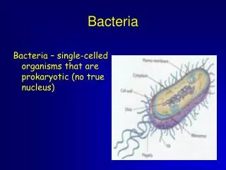
Prokaryotic Profiles: The Bacteria and the Archaea. Chapter 4. 3 Domains. Bacteria = Prokaryotic = Non-membrane bound nucleus Unicellular, but can form aggregates Found in water, soil, skin, mouth, intestine E. coli, Salmonella, Streptococcus, Staphylococcus

E N D
Prokaryotic Profiles: The Bacteria and the Archaea Chapter 4
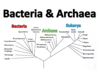
3 Domains Bacteria = Prokaryotic = Non-membrane bound nucleus • Unicellular, but can form aggregates • Found in water, soil, skin, mouth, intestine • E. coli, Salmonella, Streptococcus, Staphylococcus Archaea = Ancient prokaryotes (differ from bacteria) • Unicellular • Live in extreme environments • Methanogens (swamps, marshes, intestines) • Halophiles (salt loving) • Thermoacidophiles (hot springs & geysers) Eukarya = Eukaryotic = membrane-bound nucleus • Multicellular • Protista (Paramecium, Euglena) • Fungi (Mushrooms) • Plantae (Plants) • Animalia (Animals)
Prokaryotic Cells • Lack nucleus & membrane-bound organelles • Cell envelope • Glycocalyx • Cell wall • Plasma membrane • Cytoplasm • Nucleoid • Ribosomes • Thylakoids (cyanobacteria) • Appendages • Flagella (motility) • Sex pili (DNA transfer) • Fimbriae (attachment)
3 parts filament = long, thin, helical structure composed of proteins hook = curved sheath basal body = stack of rings firmly anchored in cell wall rotates 360o Clockwise (reverse) Counterclockwise (forward) 1-2 or many distributed over entire cell functions in motility Flagella = Bacterial Propellers
Polar flagellated cells = fastest Thiospirillum = 5.2 mm/minute Pseudomonas aeruginosa = 4.4 mm/minute Monotrichous: single flagellum at 1 end Lophotrichous: small bunches arising from 1 end of cell Amphitrichous: flagella at both ends of cell Non-polar = slowest Peritrichous: flagella dispersed over cell surface *E. coli = 1 mm/min Flagellar arrangements
Chemotaxis = move in response to chemicals • Phototaxis = move in response to light Fig 4.6
Internal flagella Axial filaments Enclosed between cell wall & cell membrane of spirochetes Provide motility twist & flex Periplasmic Flagella Fig 4.7a b
Fine hairlike bristles on cell surface Function in adhesion to other cells & surfaces Enable bacteria to attach to & infect host cells Aid in biofilm formation Fimbriae
Rigid tube made of pilin protein Longer & sparser than fimbriae Found only in Gram negative cells Functions joins bacterial cells for DNA transfer (conjugation) adhesion Pili
Glycocalyx (surface) Cell Wall (peptidoglycan) Cell Membrane (lipid bilayer) Cell Envelope3 Layers
Sticky sugar/protein coating on outside of cell 2 types capsule highly organized tightly attached slime layer loosely organized loosely attached Functions attachment inhibit phagocytosis by white blood cells aid in biofilm formation Glycocalyx
Mucoid vs Nonmucoid Bacteria • Encapsulated bacteria = generally more pathogenic due to ability to escape immune system • Streptococcus pneumoniae (pneumonia) • Bacillus anthracis (anthrax) • Haemophilus influenzae (meningitis)
Film secreted by bacteria Shower scum Plaque on teeth (Strep) Skin & mucous membranes Medical devices (Staph aureus) Storage tanks & air conditioners Biofilms = Living Layers
Cell Wall • Gives bacterial cell its shape • Surrounds & protects fragile cell membrane • Protects cell from rupturing as osmotic pressure increases • Serves as anchor point for flagella • Usually contain peptidoglycan
Peptidoglycan = a repeating framework of long glycan chains cross-linked by short peptide fragments Provides strong, flexible support to keep bacteria from bursting or collapsing due to changes in osmotic pressure Penicillin, cephalosporins, detergents, alcohol & lysozymes disrupt bonds & lyse cells Cell Wall Structure: Role of Peptidoglycan
4 Groups Based on Cell Wall Composition • Gram positive cells • Gram negative cells • Bacteria without cell walls • Bacteria with chemically unique cell walls
Gram positive Gram negative Fig 4.16
Consists of: a thick, homogenous sheath of peptidoglycan 20-80 nm thick tightly bound acidic polysaccharides teichoic acid & lipoteichoic acid negative charge controls flow of ions in & out of cell thin periplasmic space inner cell membrane Corynebacterium diphtheriae & Streptococcus express toxic proteins Retain crystal violet & stain purple Gram positive cell wall
Consists of outer membrane containing lipopolysaccharide (LPS) serves as extra barrier more difficult to kill or inhibit lipids act as endotoxins stimulate fever & shock meningitis & typhoid porins = protein channels thin peptidoglycan layer (1-3 nm) periplasmic space inner membrane Lose crystal violet & stain red from safranin counterstain Gram negative cell wall
Nontypical Cell Walls • Wall-deficient forms = Lister forms or L-phase variants • Arise from mutations in wall-forming genes • Protoplast: Gram + cell loses peptidoglycan • Spheroplast: Gram - cell loses peptidoglycan • Mycoplasmas lack cell wall • Mycobacterium have high lipid content in cell wall • waxy covering is resistant to certain chemicals & dyes
Thin (5-10nm), flexible lipid bilayer 40% phospholipids & 60% protein Mycoplasmas: high sterol content (rigid) Archaea: branched hydrocarbons Functions: Energy reactions Thylakoids in cyanobacteria Metabolic activities Nutrient processing & synthesis Transport regulation selectively permeable Secretion Cell Membrane • Mesosomes = internal folds in cytoplasm • increase surface area • aid in cell wall synthesis • prominent in Gram + bacteria
Cytoplasm • Dense gelatinous solution of sugars, amino acids & salts • 70-80% water • Serves as solvent for materials used in all cell functions • Contains: • chromatin body (DNA) • ribosomes (protein factories) • mesosomes (internal folds) • Granules (storage)
Single, circular, double-stranded DNA molecule Attached to cell membrane Contains all the genetic information required by cell DNA is aggregated in a dense area called the nucleoid Binary fission = method of prokaryotic cell division Bacterial Chromosomes
Small rings of double-stranded DNA Usually extrachromosomal Duplicated & passed to offspring Replicate independently Not essential to bacterial growth & metabolism May encode antibiotic resistance, tolerance to toxic metals, enzymes & toxins Used as vectors in genetic engineering to transfer foreign DNA Bacterial Plasmids
Site of protein synthesis Made of 60% ribosomal RNA & 40% protein Composed of large & small subunits (Svedberg units) Occur alone or in groups (polyribosomes) Free in cytoplasm or attached to membrane Prokaryotic differ from eukaryotic ribosomes in size & number of proteins Ribosomes
Intracellular storage bodies Vary in size, number & content Contents used when environmental sources are depleted glycogen poly--hydroxybutyrate (PHB) Magnetic granules (iron oxide) Gas vesicles for floating Metachromatic granules: stain contrasting color in presence of methylene blue sulfur & polyphosphate granules Corynebacterium & Mycobacterium Inclusions or Granules
Resting, dormant cell produced by G+ genera: Clostridium, Bacillus & Sporosarcina Survival mechanism, not a means for reproduction 2-phase life cycle = vegetative cell & endospore Sporulation = formation of endospores (6-8 hours) Triggered by depletion of nutrients Germination = return to vegetative growth (1.5 hours) Initiated by hydrolytic enzymes Endospores
Withstand extremes in heat, drying, freezing, radiation & chemicals thick coat high levels of calcium & dipicolinic acid (dehydrates) metabolically inactive Stringent sterilization conditions (20min @120C) Bacillus anthracis (anthrax) Clostridium tetani (tetanus) Clostridium perfringens (gangrene) Clostridium botulinum (food poisoning) Longevity verges on immortality 25-250 million years Endospores
Pleiomorphic = vary in shape and size Examples: Corynebacterium diphtheriae & mycoplasmas(lack cell walls)
Bacterial Arrangements Cocci can divide in many planes Bacilli divide only in transverse or perpendicular plane (chains or palisade)
Microscopic morphology Macroscopic morphology – colony appearance Physiological / biochemical characteristics Chemical analysis Serological analysis Genetic & molecular analysis G + C base composition DNA analysis using genetic probes Nucleic acid sequencing & rRNA analysis Methods in bacterial identification
Bergey’s Manual of Determinative Bacteriology – five volume resource covering all known procaryotes • classification based on genetic information –phylogenetic • two domains: Archaea and Bacteria • five major subgroups with 25 different phyla
Major Taxonomic Groups of Bacteria • Domain Archaea – primitive, adapted to extreme habitats and modes of nutrition • Domain Bacteria - • Phylum Proteobacteria – Gram-negative cell walls • Phylum Firmicutes – mainly Gram-positive with low G + C content • Phylum Actinobacteria – Gram-positive with high G + C content
species = a collection of bacterial cells which share an overall similar pattern of traits strain or variety = a culture derived from a single parent that differs in structure or metabolism from other cultures of that species (Serratia marcescens) type – a subspecies that can show differences in antigenic makeup (serotype), susceptibility to bacterial viruses (phage type) and in pathogenicity (pathotype) Subspecies
Tiny, gram-negative bacteria Most are pathogens that alternate between mammals and fleas, lice or ticks Intracellular pathogens Cannot survive or multiply outside of a host cell Cannot carry out metabolism on their own Rickettsia rickettisii – Rocky Mountain spotted fever Rickettsia prowazekii – epidemic typhus Coxiella burnetti – Q fever Rickettsias
Chlamydias • Require host cells for growth & metabolism • Obligate intracellular parasites • Not transmitted by arthropods • Chlamydia trachomatis – severe eye infection and one of the most common sexually transmitted diseases • Chlamydia pneumoniae – lung infections
Naturally lack a cell wall Stabilized by sterols, resistant to lysis Extremely small Range in shape from filamentous to coccus or doughnut shaped Mycoplasma pneumoniae – atypical pneumonia in humans Mycoplasmas (62,000x)
Cyanobacteria (Blue-Green) Phycocyanin = pigment Gram negative cell wall (Gracilicutes) 1 to 10 m in size Unicellular, colonial or filamentous Contain thylakoids (photosynthesis) Chlorophyll a Have gas inclusions (float) Exist in: Fresh & saltwater Hot springs Desert soil Lichens Photosynthetic Bacteria
Green & Purple Sulfur Bacteria Contain bacteriochlorophyll Utilize sulfur compounds Do not produce oxygen Anaerobic Exist in: Sulfur springs Freshwater lakes Swamps Photosynthetic Bacteria