Unraveling the DNA Structure: A Historic Journey
220 likes | 348 Views
Dive into the captivating history of DNA discovery, from Miescher's findings to the Nobel Prize-winning insights of Watson, Crick, and Wilkens. Explore the structure, composition, and significance of DNA in genetics and heredity.

Unraveling the DNA Structure: A Historic Journey
E N D
Presentation Transcript
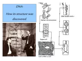
DNA- How its structure was discovered.
History of the DNA 1869- Friedrich Miescher-Swiss chemist showed that when pepsin (an enzyme that digested) proteins was used on the nucleus of cells a strange phosphorous-containing material remained. The question arose are genes composed of DNA or proteins. 1914-Robert Feulgen German chemist found a staining technique that stains more or less strongly based in the amount of DNA present. He found that all cells in an organism had the same amount of DNA except gametes, which had half the normal amount.
1928-Fred Griffith performed an experiment with 2 different strains of Pneumococcus. One was virulent and the other was not. The virulent strain had a smooth polysaccharide capsule which protected from the immune system. This allowed to caused pneumonia in mice and killed them. The other strain did not have the capsule and was "rough". This strain could not cause pneumonia in mice. When Griffith injected the rough strain of bacteria in mice they lived, and when the smooth strain of bacteria was injected into the mice they died. He killed some of the smooth bacteria by heating them and then injecting them into the mice. The mice lived. He then took some of the killed smooth bacteria and some of the rough bacteria and mixed them together. This bacteria then had the ability to kill mice. This is because the rough bacteria had been "transformed" by taking up some of the DNA from the smooth bacteria.
The conclusion was that the bacteria had incorporated heredity from source and in doing so expressed a new smooth trait. 1944-Avery, McLeod and McCarty tried mixing the rough strain with different chemical from the S strain and it was found the DNA extracted from the smooth-strain and transform the rough strain.
Alfred Hershey and Martha Chase demonstrated the genetic material is DNA by using viruses that infect bacteria. These viruses only stay on the outside of the cell when infecting. Also viruses are made of protein and DNA.
It demonstrated that DNA is the material that genes are made of and not protein.
Chargaff's Rule and that A+G=C+T=50% A T G C Humans 30.9 29.4 19.9 19.5 Wheat 27.3 27.1 22.7 22.8 Yeast 31.3 32.9 18.7 17.1 Sarcina lutea 13.4 12.4 37.1 37.1 T7 26.3 26.0 24.0 24.0
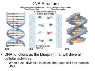
Wilkens and Franklin used x-ray crystallography to determine that DNA was double stranded, a helix, phosphates were on the outside and three distances, 2.0 nm, .34 nm, and 3.4 nm showed up in a pattern over and over again
Watson and Crick determined that 2.0 nm was the distance from one strand to the other. .34 nm was the distance from one base pair to another and finally 3.4 nm determined that there were 10 bases to a complete twist in the helix. So with 2.0 nm from one strand to the other, it was determined that the a purine had had to be base paired with a pyrimidine. Then looking at the hydrogen bonding, adenine base paired with thyamine because they could form two hydrogen bonds and guanine base paired with cytosine because they could form three hydrogen bonds. Watson, Crick, and Wilkens received the Nobel Prize in 1962. Unfortunately, Franklin died of cancer at the age 38.
Two purines are too wide and would overlap. Two pyrimidines are too far apart to form the hydrogen bonds. A purine and a pyrimidines however, are just right!
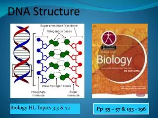
DNA is the longest molecule found in the cell, yet its structure is quite simple. The human cell contains 5-6 feet of DNA in every cell autosomal cell. The basic building block of nucleic acids are the nucleotides. There are 3 billion base pairs or 6 billion nucleotides in a human cell. A nucleotide consist of 1. A pentose sugar Ribose for RNA and Deoxyribose for DNA 2. Phosphate group- This is what makes the nucleic acid, acidic 3. Nitrogenous base- DNA- AGCT RNA- AGCU
DNA is double stranded and analogous to a ladder. The sides of the ladder are composed of alternating sugars (deoxyribose) and phosphate groups that run antiparallel to one another. On the left side (next card) the first carbon found on the strand is #5 and moving on down the last carbon is carbon # 3. This side is said to be 5'-3'. The opposite side is upside down compared to the other side. The right hand side, the first carbon found on the strand is #3 and moving on down the last carbon is carbon # 5. This side is said to be 3'-5'.
The nitrogenous bases form the rungs of the ladder. Thyamine will base pair with adenine on the opposite side. This is a pyrimidine base paired with a purine. This will form 2 hydrogen bonds. Hydrogen bonds are weak but millions of them together will keep the two strands together.
Guanine will base pair with cytosine on the opposite side. This is a pyrimidine base paired with a purine. This will form 3 instead of 2 hydrogen bonds.
This will continue for billions of base pairings forming a molecule of DNA.