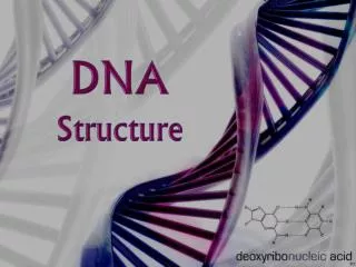
DNA STRUCTURE
Dive into the intricate world of DNA structure with Dr. Amena Rahim's expert insights. Nucleic acids, vital for genetic information storage, come in two forms: DNA and RNA. Discover how DNA, found in chromosomes and organelles, exists primarily as a double helix. Learn about covalent 3′→5′ phosphodiester bonds that link nucleotides, the significance of base pairing, and the role of major and minor grooves. Explore the implications of DNA structure in gene expression and health, including how specific drugs target DNA. Ideal for students and enthusiasts in biochemistry.

DNA STRUCTURE
E N D
Presentation Transcript
DNA STRUCTURE DR AMENA RAHIM BIOCHEMISTRY
Nucleic acids are required for the storage and expression of genetic information. • There are two chemically distinct types of nucleic acids: deoxyribonucleic acid (DNA) and ribonucleic acid
DNA, the repository of genetic information, is present in chromosomes in the nucleus of eukaryotic organisms, • in mitochondria and • the chloroplasts of plants.
Prokaryotic cells, which lack nuclei, have a single chromosome, but may also contain nonchromosomal DNA in the form of plasmids.
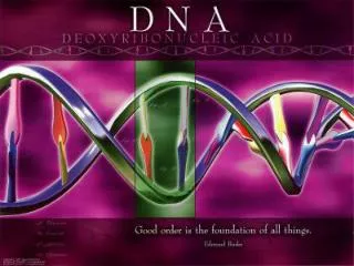
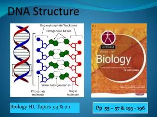
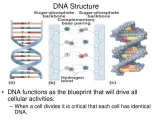
Structure of DNA • DNA is a polymer of deoxyribonucleosidemonophosphates covalently linked by 3′→5′–phosphodiester bonds. • With the exception of a few viruses that contain single-stranded (ss) DNA, DNA exists as a double-stranded (ds) molecule, in which the two strands wind around each other, forming a double helix.
In eukaryotic cells, DNA is found associated with various types of proteins (known collectively as nucleoprotein) present in the nucleus, • whereas in prokaryotes, the protein–DNA complex is present in a nonmembrane-bound region known as the nucleoid.
3′→5′-Phosphodiester bonds • Phosphodiester bonds join the 3′-hydroxyl group of the deoxypentose of one nucleotide to the 5′-hydroxyl group of the deoxypentose of an adjacent nucleotide through a phosphate group.
The resulting long, unbranched chain has polarity, with both a 5′-end (the end with the free phosphate) and a 3′-end (the end with the free hydroxyl) that are not attached to other nucleotides.
The bases located along the resulting deoxyribose–phosphate backbone are, by convention, always written in sequence from the 5′-end of the chain to the 3′-end.
Phosphodiester linkages between nucleotides (in DNA or RNA) can be cleaved hydrolytically by chemicals, or hydrolyzed enzymatically by a family of nucleases: deoxyribonucleases for DNA and ribonucleases for RNA.
Double helix • In the double helix, the two chains are coiled around a common axis called the axis of symmetry. The chains are paired in an antiparallel manner, that is, the 5′-end of one strand is paired with the 3′-end of the other strand.
In the DNA helix, the hydrophilic deoxyribose–phosphate backbone of each chain is on the outside of the molecule, whereas the hydrophobic bases are stacked inside.
The spatial relationship between the two strands in the helix creates a major (wide) groove and a minor (narrow) groove.
These grooves provide access for the binding of regulatory proteins to their specific recognition sequences along the DNA chain. • Certain anticancer drugs, such as dactinomycin (actinomycin D), exert their cytotoxic effect by intercalating into the narrow groove of the DNA double helix, thus interfering with DNA and RNA synthesis.
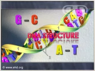
Base pairing • The bases of one strand of DNA are paired with the bases of the second strand, so that an adenine is always paired with a thymine and a cytosine is always paired with a guanine. • The base pairs are perpendicular to the axis of the helix.
The specific base pairing in DNA leads to the Chargaff Rule: In any sample of dsDNA, the amount of adenine equals the amount of thymine, the amount of guanine equals the amount of cytosine, and the total amount of purines equals the total amount of pyrimidines.
The base pairs are held together by hydrogen bonds: two between A and T and three between G and C . • These hydrogen bonds, plus the hydrophobic interactions between the stacked bases, stabilize the structure of the double helix.
Separation of the two DNA strands in the double helix • The two strands of the double helix separate when hydrogen bonds between the paired bases are disrupted. • Disruption can occur in the laboratory if the pH of the DNA solution is altered so that the nucleotide bases ionize, or if the solution is heated.
Phosphodiester bonds are not broken by such treatment. • When DNA is heated, the temperature at which one half of the helical structure is lost is defined as the melting temperature (Tm).
The loss of helical structure in DNA, called denaturation, can be monitored by measuring its absorbance at 260 nm. • ssDNA has a higher relative absorbance at this wavelength than does dsDNA.
Because there are three hydrogen bonds between G and C but only two between A and T, • DNA that contains high concentrations of A and T denatures at a lower temperature than G- and C-rich DNA.
Under appropriate conditions, complementary DNA strands can reform the double helix by the process called renaturation (or reannealing).
Structural forms of the double helix: • There are three major structural forms of DNA: the B form, described by Watson and Crick in 1953. • the A form • the Z form.
The B form is a right-handed helix with ten residues per 360° turn of the helix, and with the planes of the bases perpendicular to the helical axis. Chromosomal DNA is thought to consist primarily of B-DNA.
The A form. It is also a right-handed helix, but there are eleven base pairs per turn, and the planes of the base pairs are tilted 20° away from the perpendicular to the helical axis. • The conformation found in DNA–RNA hybrids or RNA–RNA double-stranded regions is probably very close to the A form.
Z-DNA is a left-handed helix that contains about twelve base pairs per turn . • Stretches of Z-DNA can occur naturally in regions of DNA that have a sequence of alternating purines and pyrimidines, for example, poly GC.
Transitions between the helical forms of DNA may play a role in regulating gene expression.
Circular DNA molecules • Each chromosome in the nucleus of a eukaryote contains one long linear molecule of dsDNA, which is bound to a complex mixture of proteins to form chromatin.
Eukaryotes have closed circular DNA molecules in their mitochondria, as do plant chloroplasts. • A prokaryotic organism contains a single, double-stranded, supercoiled, circular chromosome. Each prokaryotic chromosome is associated with histone-like proteins and RNA that can condense the DNA to form a nucleoid.
Most species of bacteria also contain small, circular, extrachromosomal DNA molecules called plasmids. • Plasmid DNA carries genetic information, and undergoes replication that may or may not be synchronized to chromosomal division.
Plasmids may carry genes that convey antibiotic resistance to the host bacterium, and may facilitate the transfer of genetic information from one bacterium to another.
Organization of Eukaryotic DNA • A typical human cell contains 46 chromosomes, whose total DNA is approximately 1m long! • DNA interacts with a large number of proteins, each of which performs a specific function in the ordered packaging of these long molecules of DNA.
Eukaryotic DNA is associated with tightly bound basic proteins, called histones. These serve to order the DNA into basic structural units, called nucleosomes, that resemble beads on a string.
Nucleosomes are further arranged into increasingly more complex structures that organize and condense the long DNA molecules into chromosomes that can be segregated during cell division. • The complex of DNA and protein found inside the nuclei of eukaryotic cells is called chromatin.
Histones and the formation of nucleosomes • There are five classes of histones, designated H1, H2A, H2B, H3, and H4. • These small proteins are positively charged at physiologic pH as a result of their high content of lysine and arginine. Because of their positive charge, they form ionic bonds with negatively charged DNA.
Histones, along with positively charged ions such as Mg+2, help neutralize the negatively charged DNA phosphate groups.
Nucleosomes: • Two molecules each of H2A, H2B, H3, and H4 form the structural core of the individual nucleosome “beads.” • Around this core, a segment of the DNA double helix is wound nearly twice, forming a negatively supertwisted helix.
The N-terminal ends of these histones can be acetylated, methylated, or phosphorylated. • These reversible modifications can influence how tightly the histones bind to the DNA, thereby affecting the expression of specific genes.