Kidneys and Urination
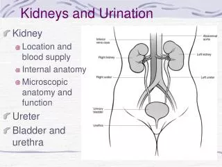
Kidneys and Urination. Kidney Location and blood supply Internal anatomy Microscopic anatomy and function Ureter Bladder and urethra. Location of kidney. Retroperotoneal at mid-abdomen T12-L3 NAV enters at hilus Renal aa. off aorta Renal vv to IVC Nerves all autonomics--renal plexus

Kidneys and Urination
E N D
Presentation Transcript
Kidneys and Urination • Kidney • Location and blood supply • Internal anatomy • Microscopic anatomy and function • Ureter • Bladder and urethra
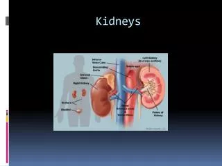
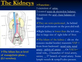
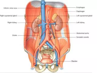
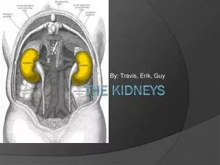
Location of kidney • Retroperotoneal at mid-abdomen • T12-L3 • NAV enters at hilus • Renal aa. off aorta • Renal vv to IVC • Nerves all autonomics--renal plexus • Ureter--exits at hilus • “Ad”renal gland superior to kidney--unrelated in function • Own blood supply • Endocrine gland
Internal anatomy of kidney • NAV branch out from hilus • Collecting ducts unite and urine leaves through ureter at hilus • Cortex is outer/superficial tissue • Light, granular • Functioning nephrons here • Medulla is inner/deep tissue • Darker • Pyramid-cone shape • Collecting tubules unite into ducts into ureter
Internal anatomy of kidney--details • Lobe of kidney is medullary pyramid plus cortex around it • Cortex contains urine-concentrating nephrons • Medullary pyramids • Tubules receive concentrated urine from cortex • Appear striated because contains parallel converging urine-collecting tubules • Flow of urine • Collecting tubes of medullary pyramid minor calyx major calyx renal pelvis ureter Pg 679
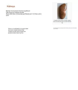
Microscopic anatomy and function • Nephron or urine-concentrating unit is in outer cortex--millions • Capillaries surround glomerulus (ball) and filtrate passes into duct • Counter-current exchange in ducts (Loop of Henle) concentrates urine) More details at “How Stuff Works” http://science.howstuffworks.com/kidney.htm/printable
Ureter--from kidney to bladder LAYERS OF URETER • External connective tissue--adventitia • Middle muscular layer--muscularis • Smooth Muscle • Inner Longitudinal • Outer Circular • External longitudinal (on distal third) • Peristaltic action moves urine to bladder (and stones!!) • Inner lining of transitional (stretchy) epithelium--Mucosa
Bladder • Muscular (what kind?) sac that fills with urine from ureters • Anterior against pubis in pelvis (more with pelvis) • Filled with urine expands into abdomen • Blood supply from internal iliac arteries • Innervation is autonomic from hypogastric plexus
Layers of bladder wall • Outer connective tissue--adventitia • Middle muscular layer (“detrusal” or expulsor)--inner and outer longitudinal fibers around middle circular fibers • Inner transitional (stretchy) epithelium • Bladder can expand 15 times its empty volume to hold 500 ml of urine • Trigone is triangle between ureters/urethra--persistent sight of infection
Urethra • Drains urine from bladder to outside • Female = short tube • Males = long tube • Prostatic, Membranous, Spongy (penile) portions • Also carries sperm • Internal Urethral Sphincter • Between bladder + urethra • Thickening of detrusor (smooth muscle) • External Urethral Sphincter • Within urogenital diaphragm • Skeletal muscle = voluntary control urination • External Urethral Orifice • Males = end of penile urethra • Females = anterior to vaginal opening, posterior to clitoris (more later with pelvis)
Micturition = Urination • Emptying bladder • Stretch receptors in bladder respond when bladder full • Parasympathetic signals detrusor muscle to contract and internal urinary sphincter to open (also inhibits sympathetic pathways that would prevent urination) • Other brain receptors can inhibit urination by relaxing detrusor, and keep external urinary sphincter closed • Voluntary contraction of abdominal wall muscles increases abdominal pressure • Voluntary relaxation of external urethral sphincter For nice review of kidneys and urination http://webanatomy.net/anatomy/urinary_notes.htm See pg 692, M&M
Ascent of the kidney in development • Kidneys from intermediate mesoderm • Pronephric kidney in fetus shows segmental body plan • Fish with dorsal renal tissue lateral to vertebral column for most of length • In human, metanephric kidney migrates from inferior to superior • Variation in kidney shape not uncommon (horseshoe kidney • Ureter also from intermediate mesoderm