Kidneys
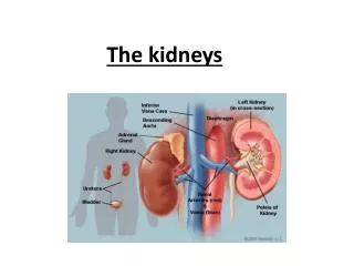
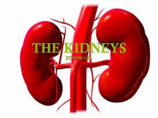
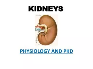
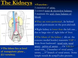
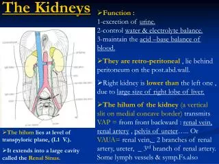
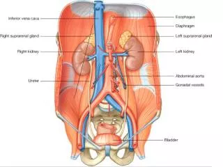
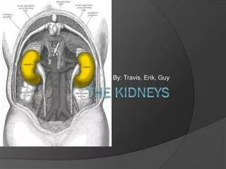
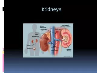
Kidneys. What do kidneys do?. What do the kidneys do? The kidneys are bean-shaped organs each about the size of a fist located near the middle of the back, just below the rib cage, one on each side of the spine.

Kidneys
E N D
Presentation Transcript
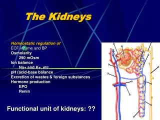
What do kidneys do? What do the kidneys do? • The kidneys are bean-shaped organs • each about the size of a fist • located near the middle of the back, just below the rib cage, one on each side of the spine. • Every day, a person’s kidneys process about 200 quarts of blood to sift out about 2 quarts of waste products and extra water. • The wastes and extra water become urine, which flows to the bladder through tubes called ureters. • The bladder stores urine until releasing it through urination. • Wastes in the blood come from the normal breakdown of active tissues, such as muscles, and from food. • The body uses food for energy and self-repairs. • After the body has taken what it needs from food, wastes are sent to the blood. • If the kidneys did not remove them, these wastes would build up in the blood and damage the body.
What is a nephron • The actual removal of wastes occurs in tiny units inside the kidneys called nephrons. • Each kidney has about a million nephrons. • In the nephron, a glomerulus—which is a tiny blood vessel, or capillary—intertwines with a tiny urine-collecting tube called a tubule. • The glomerulus acts as a filtering unit, or sieve, and keeps normal proteins and cells in the bloodstream, allowing extra fluid and wastes to pass through. • A complicated chemical exchange takes place, as waste materials and water leave the blood and enter the urinary system.
ureters • Two ureters - narrow tubes that carry urine from the kidneys to the bladder. • Muscles in the ureter walls continually tighten and relax forcing urine downward, away from the kidneys. I • f urine backs up, or is allowed to stand still, a kidney infection can develop. • About every 10 to 15 seconds, small amounts of urine are emptied into the bladder from the ureters.
bladder • bladder • a triangle-shaped, hollow organ located in the lower abdomen. • It is held in place by ligaments that are attached to other organs and the pelvic bones. • The bladder's walls relax and expand to store urine, and contract and flatten to empty urine through the urethra. • two sphincter muscles - circular muscles that help keep urine from leaking by closing tightly like a rubber band around the opening of the bladder. • nerves in the bladder - alert a person when it is time to urinate, or empty the bladder.
urethra • the tube that allows urine to pass outside the body. • The brain signals the bladder muscles to tighten, which squeezes urine out of the bladder. • At the same time, the brain signals the sphincter muscles to relax to let urine exit the bladder through the urethra. • When all the signals occur in the correct order, normal urination occurs.
FYI Facts about urine: • Adults pass about a quart and a half of urine each day, depending on the fluids and foods consumed. • The volume of urine formed at night is about half that formed in the daytime. • Normal urine is sterile. It contains fluids, salts and waste products, but it is free of bacteria, viruses and fungi. • The tissues of the bladder are isolated from urine and toxic substances by a coating that discourages bacteria from attaching and growing on the bladder wall.
Nephron structure • Kidney nephrons are the functional units of the kidneys. There are typically over 10,000 kidney nephrons in each of the two kidneys in the body.
renal cortex • The cortex is the outer part of the kidney. • This is where blood is filtered. • Billions of glomeruli are found in the cortex. • A glomerulus is a tiny ball of capillaries. • Each glomerulus is surrounded by a "Bowman's Capsule". • Things like red blood cells, white blood cells, platelets and fibrinogen stay in the blood vessels.
renal medulla • The medulla is the inside part of the kidney. • This is where the amount of salt and water in your urine is controlled. • It consists of billions of loops of Henlé. • These work very hard pumping sodium ions. • . • The opposite of an anti-diuretic is a "diuretic". Alcohol and tea are diuretics.
glomerus • the glomerulus filters proteins and cells from the blood.
collecting ducts Collecting ducts run through the medulla and are surrounded by loops of Henlé. It is called a collecting duct because it collects the liquid produced by lots of nephrons.
Bowman capsule A cup-shaped structure around the glomerulus of each nephron is called the Bowman Capsule. It serves as a filter to remove organic wastes, excess inorganic salts, and water. Bowman's capsule is named after its identifier, English physician and physiologist, Sir William Bowman (1816-1892).
URINARY SYSTEM SIGNS, SYMPTOMS, & DISEASES
azoturia • Increase of nitrogen substances, especially urea in urine. • Versus azotemia or uremia = when it happens in the blood
diuresis • Diuresis is an increase in the production and secretion of urine • Diuretics- are meds that increase the amount of urine a person makes
dysuria • refers to painfulurination • This is typically described to be a burning or stinging sensation. • It is most often a result of a urinary tract infection. • It may also be due to an STD, bladder stones, bladder tumours, and virtually any condition of the prostate.
end stage renal disease (ESRD) • End-stage kidney disease is the complete, or almost complete failure of the kidneys to function. • The kidneys can no longer adequately filter the blood. • Ultimately requires dialysis or transplantation to survive. • CRF – Chronic renal failure • Diabetes the most common cause.
enuresis • Involuntary discharge of urineafter the age (5) when bladder control should be established. • Nocturnal enuresis, commonly called bedwetting, is a medical term for involuntary urination while asleep after the age at which bladder control usually occurs
hypospadias • Abdnormalcongenital opening of the male urethra on the undersurface of the penis. • Instead of opening at the tip of the glans of the penis, a hypospadic urethra opens anywhere along a line (the urethral groove) running from the tip along the underside (ventral aspect) of the shaft to the junction of the penis and scrotum or perineum
interstitial nephritis • A pathological change in the renal interstitial tissue. Results in destruction of the nephrons and renal impairment. • Due to toxic agent like drugs or chemicals. • This disease can be either acute, meaning it occurs suddenly, or chronic, meaning it is ongoing and eventually ends in kidney failure.
renal hypertension • High blood pressure that results from kidney disease.
uremia • Elevated levels of urea and other nitrogen waste products in the blood • Also called azotemia • In kidney failure, urea, nitrogenous waste products , and other waste products, which are normally excreted into the urine, are retained in the blood.
Wilms tumor • Maligant neoplasm of the kidneys that typically occurs in children before age 5, rarely in adults • Signs – hypertension, palpable mass, and hematuria
URINARY SYSTEM DIAGNOOSTIC PROCEDURES
blood urea nitrogen (BUN) Laboratory test is a measure of the amount of urea in the blood (nitrogen in the blood in the form of urea) and a measurement of the kidneys’ ability to filter urea from the blood Urea is a substance secreted by the liver, and removed from the blood by the kidneys. Increase BUN can mean impaired kidney function
CT Scan Computed tomography • Radiographic exam • Full arc around patient to produce cross-sectional images or slices
kidney, ureter, bladder (KUB) • Radiographic exam • Determines location, shape, and malformation of the KUB
PYELOGRAPHYRadiographic study of KUB with an injection of a contrast • IVP- Intavenouspyelography • Contrast injected intravenously • Visualize the ENTIRE urinary tract • Also called IVU (urography) and EU (excretory urography) • RP- Retrograde Pyelography • Contrast injected through a cystoscope into the bladder and ureters by a catheter. • Details collection system of the kidney • Shows obstructions
RENAL SCAN • Nuclear medicine image • Determines renal function and shape • Radioactive substance injected IV and concentrates in the kidney
URINALYSIS (UA) • Evaluation of the urine • Physical • Chemical • Microscopic
VOIDING CYSTOURETHROGRAPHY (VCUG) • Radiography of the bladder and urethra • After filling the bladder with a contrast • And during the process of voiding (peeing)
URINARY SYSTEM MEDICAL AND SURGICAL PROCEDURES
CATHETERIZATION • Insertion of a catheter (hollow flexible tube) into a body cavity or organ • Instills a substances or removes fluid • Typically through the urethra into the bladder to drain urine
DIALYSIS HEMODIALYSIS • Remove excess fluids and toxins from the blood by shunting • Taking the patient’s blood from the body into a dialysis machine where it is filtered, cleaned and returned to the body • See page 310
DIALYSIS PERITONEAL DIALYSIS • Using the patient’s own peritoneum (space in the abdomen) as the dialysis membrane. • Can be continuous or interminttent. • See page 311
RENAL TRANSPLANTATION • Organ transplant of a kidney • A person with ESRD • Also called a kidney transplant