Download
1 / 1
20 likes | 128 Views
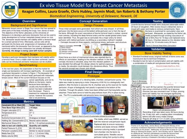
Ex vivo Tissue Model for Breast Cancer Metastasis. Keagan Collins, Laura Graefe , Chris Hubley , Jaymin Modi, Ian Roberts & Bethany Porter Biomedical Engineering, University of Delaware, Newark, DE. Overview. Concept Generation. Testing.

E N D
Ex vivo Tissue Model for Breast Cancer Metastasis Keagan Collins, Laura Graefe, Chris Hubley, Jaymin Modi, Ian Roberts & Bethany Porter Biomedical Engineering, University of Delaware, Newark, DE Overview Concept Generation Testing Juvenile bovine femoral heads were analyzed (obtainable within 24 hours of slaughter). After excess tissue is detached, bone is rinsed with water and media to remove contaminants. Next, Background and Significance Design Process Metastatic breast cancer is Stage Four cancer that has spread to other parts of the body including the lungs, liver, bones or brain. The objective of the Nohe Laboratory at the University of Delaware is to develop a perfusion bioreactor that can be used to study development of human metastatic breast cancer ex vivo. The bioreactor would maintain cell viability within a femoral head for a certain period of time. Afterwards, cancer cells will be perfused into the bone and progression of metastasis will be monitored within the bioreactor from the onset, as opposed to the commonly models where metastasis has already developed.Currently, the design is being used with human samples. • Three initial concepts were generated, from which one was chosen. In all three, perfusion into the bone occurs at the bottom while perfusion out is from the top of the bone. Although the exact vasculature of bovine femoral head is unclear, several main arteries, including the deep femoral artery and the caudal gluteal artery have been identified. Thus, the goal was to cannulate into a major artery for direct perfusion with indirect perfusion via drilling as a secondary method if needed. • Concepts focused on three aspects: mechanical stress, perfusion, and structure. the bone is examined for cannulation sites and perfused. Afterwards, as stated by the Nohe Lab, media pumped through existing vasculature at rate of 8mL/hour for 10 days and replaced every 36 hours. Chamber placed on the vibration table (60 Hz) for 10 minutes every 24 hours Figure 5: Femoral Head Artery Cannulation Figure 2: Concepts [1] Direct Stress [2] Vibration Stress [3] Vibration and Elevation [4] Final Concept Validation 2 3 1 Bone Viability Testing Project Scope • Standard live/dead assay (original method) of bone cores was inconclusive due to technical limitations. • Decided to test for lack of contamination and cell viability with a combination of media pH and glucose level monitoring. Develop a perfusion bioreactor that maintains cell viability within a femoral head. Once a viable state has been achieved, cancer cells can be introduced and bone metastasis can be studied. Direct force as a form of mechanical stress was rejecteddue to effects on cannulation, leading to the vibration method. In the third concept, the bone is elevated, away from the vibration platform. It was decided that although this design allows for optimal exposure to media, the elevation could dampen the vibrational effects. Thus the second concept was chosen and refined. 4 Benchmarking In the next ten years, the expectation is that the market for single use bioreactors will increase significantly. An example of a perfusion bioreactor is shown in Figure 1.The bioreactor for this project will house a femoral head, perfused with metastatic breast cancer. Most of the current literature focuses on the . Final Design Prototype • The final design consists of a vibrating table, chamber, and perfusion pump. The table was made by epoxying a motor (frequency of 60 Hz) to a biologically inert platform. After the bone is secured, the 4L chamber acts as the reservoir for media perfusion. A layer of biologically inert plastic is epoxied to the bottom of the chamber. Through this plastic, holes have been drilled such that long bolts can be attached. These bolts serve to hold the polyurethane foam (PU) in place during use. The PU is autoclavable and prevents bone movement during vibration. cancer specifically, and not its effects on the bone, mainly because bone damage is usually only found after the cancer has been eradicated from the breast. There is treatment for metastatic breast cancer, however the success rate is not promising Figure 6: Microscopy of cell waste products, - For each 36 hour period, the juvenile femoral head was using a minimum of 4300 mg of glucose. - Glucose use was consistent over 10 days, proving bone cell viability has not changed significantly. - No visible media contamination (bacterial or other) Figure 1: Perfusion Bioreactor Metrics Figure 7: pH and glucose testing - Recording of pH was taken every 36 hours during the 10 day trial, with a reading taken immediately before and after every media change. - Regular pH drop of 1.0-1.5 (cells using nutrients). Figure 3: final 2-D and 3-D schematics followed by prototype The media, which was DMEM, served as both the reservoir and perfusion liquid. Replaced daily to ensure necessary nutrients and waste removal, DMEM (common culturing media) is pumped from chamber into the bone via cannulated points. Needles secure perfusion tubing from pump to artery. Acknowledgements Thanks to Dr. AnjaNohe, Jeremy Bonor and HemanthAkkiraju of the University of Delaware Department of Biological Sciences for their support and funding for the project. Additionally, we would like to thank Dr. Liyun Wang for advising the project, and Dr. Jenni Buckley and Dr. Robert Sikes for additional aid. Herman’s delicatessen provided the femoral head samples that were used for this project. Lastly, we would like to acknowledge University of Delaware’s Biomedical Engineering Department, Figure 4: cannulation technique