Download

1 / 64
650 likes | 812 Views
Screening for Breast cancer. The Obstetrics & Gynecological Society of Bhopal & AMPOGS Research Public Welfare Society. Screening tools. Clinical Breast examination Breast self examination Mammography Ultrasonography/ elastography FNAC Cytology of nipple discharge.

E N D
Screening for Breast cancer The Obstetrics & Gynecological Society of Bhopal & AMPOGS Research Public Welfare Society
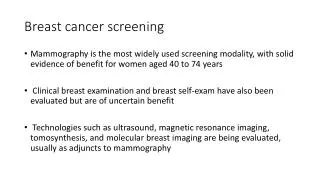
Screening tools • Clinical Breast examination • Breast self examination • Mammography • Ultrasonography/elastography • FNAC • Cytology of nipple discharge
AGE STANDARDISED (WORLD) BREAST AND GENITAL TRACT CANCER INCIDENCE RATES PER 100,000 FEMALES 35 31.3 30 28.2 27.5 24.6 23.3 25 23.2 20.2 19.3 20 20.1 21.2 19.3 15.7 17.4 16.6 15 RATE 10 7.6 7.2 7.2 7.2 8.3 6.5 4.8 5 2.3 2.5 2.4 3.2 1.6 1.4 1.3 0 1970 1975 1980 1985 1990 1995 2000 YEAR CERVIX UTERI OVARY CORPUS UTERI BREAST
Breast cancer Incidence • Most common cancer in women worldwide. • Most common cause of death from cancer among women. • More than three fourths of these women in developing countries are diagnosed in advanced stage of the disease. If these lesions are detected early, most breast cancers can be effectively treated with good outcome. • In India 144,937 women were newly detected with breast cancer in 2012, of which 70,218 women died. Roughly, for every 2 women newly diagnosed with breast cancer in India, one dies of this disease.
Who to be screened • Women between the ages of 40-60 years of age • All women identified with a breast mass that has previously not been clinically evaluated need to be screened for breast cancer • Women with high Risk factors can be offered screening from age 30 years such as • Age over 40 • No children or children after 30 years of age • Mother or sister with breast cancer • History of breast biopsies or breast cancer • Initiation of menses before 12 years of age • Overweight • Screening to be every 2 years
Clinical Breast examination - Tips • Be sensitive to the woman by giving her opportunities to express any concerns before and during the examination. • Respect the woman’s sense of privacy. • If the woman is anxious, assure her that you will do your best to make the examination comfortable. • Throughout the examination, approach the woman slowly and avoid any sudden or unexpected movements. • Do not rush through the examination. Perform each step gently and ask her if she is having any discomfort during any part of the examination. Be aware of her facial expressions and body movements as indications that she is uncomfortable. • Always take into consideration any cultural factors when deciding what clothing the woman should remove. Have a clean sheet or drape to cover the woman’s breast if needed. • These examinations should be performed in a clean, well-lit, private examination or procedure room that has a source of clean water. A female assistant should be available to accompany the woman when a male clinician is the examiner.
Getting ready • Tell the woman you are going to examine her breasts. • This is a good time to ask if she has noted any changes in her breasts and whether she does monthly breast self-examinations. Tell the woman that you will show her how to do a breast self-examination before she leaves. • Wash your hands thoroughly with soap and water and dry them with a clean, dry cloth or allow them to air dry before beginning the examination. • If there are open sores or nipple discharge, put new examination or high-level disinfected surgical gloves on both hands. • Ask the woman to undress till the waist. With the woman undressed from the waist up, have her sit on the examining table with her arms at her sides. • Examine both in sitting and lying down position
Performing a CBE • Steps of examination - CBE involves two main parts: • Inspection to identify physical signs of breast cancer. • Palpationwhich involves using the finger pads to physically examine all areas of breast tissue including lymph nodes (underarm area) to identify lumps • 4 positions • Arms by the side of trunk. • Raising arms over the head. • Pressing on the hips. • Leaning forward.
inspection • In the sitting position first visually inspect the breast, initially when woman is sitting up right with arms on her hips, and then with her arms raised over head. • Note any change in symmetry of breast shape, size, skin changes–skin dimpling or retraction or ulceration the level of both nipples, retraction of nipple(s), inverted nipple.
Look at the breasts for shape and size. • Note any difference in shape, size, nipple or skin puckering or dimpling. Although some difference in size of the breasts is normal, irregularities or difference in size and shape may indicate masses. • Swelling, increased warmth or tenderness in either breast may suggest infection, especially if the woman is breastfeeding.
Look at the nipples and note their size and shape and the direction in which they point (e.g., do her breasts hang evenly?). Also check for rashes or sores and any nipple discharge. • Have the woman first raise her arms over her head and then press her hands on her hips to contract her chest wall (pectoral) muscles. In each position, inspect the size, shape and symmetry, nipple or skin puckering or dimpling of the breast and note any abnormalities. (These positions will also show skin puckering or dimpling if either is present.) Then have the woman lean forward to see if her breasts hang evenly.
palpation • Have the woman lie down on the examining table. • Placing a pillow under her shoulder on the side being examined will spread the breast tissue and may help in examining the breast. • Place a clean sheet or drape over the breast you are not examining. • Place the woman’s left arm over her head. Look at the left breast to see if it looks similar to the right breast and whether there is puckering or dimpling. • Use “Dial of clock method” for palpation, first use the finger pads of the middle three fingers to palpate the entire breast, in overlapping circular motions, one area at a time. Repeat both parts of the examination on both the left and rights breasts.
Woman in lying down position Palpation pressure • Light pressure for superficial breast tissue • Medium pressure for intermediate layer • Deep pressure for tissue close to chest wall • The finger pads of middle three fingers should be used to palpate the breast in circular motion
PALPATION Pads of three middle fingers, hand bowed up Dime size circles JAMA, Vol. 282, No 13, Oct. 1999 Slide between palpations without lifting fingers
Spiral technique • Using the pads of your three middle fingers, palpate the breast using the spiral technique. Start at the top outermost edge of the breast. Press the breast tissue firmly against the ribcage as you complete each spiral and gradually move your fingers toward the areola. Continue this until you have examined every part of the breast. Note any lumps or tenderness.
Check for nipple discharge • Using the thumb and index finger, gently squeeze the nipple of the breast. Note any discharge: clear, cloudy or bloody. Any cloudy or bloody discharge expressed from the nipple should be noted in the woman’s record. Although it is normal to have some cloudy discharge from either or both breasts up to a year after giving birth or stopping breastfeeding, rarely it may be due to cancer, infection or a benign tumor or cyst. Repeat these steps for the right breast.
Axillary tail/ lymph nodes • To palpate the tail of the breast, have the woman sit up and raise her left arm to shoulder level. If needed, have her rest her hand on your shoulder. Press along the outside edge of the pectoral muscle while gradually moving your fingers up into the axilla to check for enlarged lymph nodes or tenderness. It is essential to include the tail of the breast in the palpation because this is where most cancer occurs.
Dial of a clock method Palpation will be done in each segment until entire breast is covered. • Pads of finger (not tips of fingers) of middle three fingers (index, middle and ring) with hand held in slightly bowed position will be used for palpation. • In the “dial of a clock” method the whole breast is palpated as if it was a dial of a clock, 12 O’ clock being the highest point at upper edge of breast just below the midclavicular point and 6 O’ clock being at the inframammary crease. The palpation is begun at 12 O’clock from periphery to the nipple by describing small circles of about 3 cm in diameter. Following circular movement of the “pad of fingers” 3 times with increasing pressure and without lifting the fingers, the next circle is felt towards the nipple , overlapping with the previous circle to about half in diameter. Once the areola and nipple area is reached, the next segment /sector is palpated at 1-O’clock. The procedure of palpation with “pad of 3 fingers” is repeated sequentially at 2 0’ clock, 3 0’, 4 0’, 5 0’, 6 0’, 7 0’, 8 0’, 9 0’, 10 0’ and 11 0’. If a lump is detected, its size should be measured using a Vernier caliper. The palpation of mammary ducts is done by gently rolling the ducts between the index finger and the thumb. Any thickening, tenderness or discharge is noted while palpating the mammary ducts. In case of retraction of the nipple an attempt is made to pull the nipple forward to see if the nipple could be brought forward or not and if any lump is present underneath the areola, whether the nipple and the ducts are tethered to the lump or not. The skin overlying the lump is gently pinched and moved with the fingers to see if the skin could be moved freely from /off the lump. If the skin is free from the lump but the movement of lump away from skin causes dimpling of skin, the skin is considered “tethered”. If no movement of skin is possible, it is considered “fixed”. The fixity of lump to underlying pectoralis major muscle is ascertained by requesting the lady to push her hand against the hip to contract the muscle and then moving the lump.
Note any discharge from the nipple(s), colour of the discharge, swelling/ lumps, consistency of the lumps, swelling in the armpit (axillary area), above the collar bone (supraclavicluar area) and root of the neck (infraclavicular area). • Repeat this step for the right side. • After completing the examination, have the woman dress herself. Explain any abnormal findings and what, if anything, needs to be done. If the examination is entirely normal, tell her everything is normal and healthy and when she should return for a repeat examination (i.e., annually or if she finds any changes on breast self-examination). • The optimal time for a CBE in a premenopausal woman is 5-10 days after the onset of menses, avoiding the week before the period is preferable. Postmenopausal women may have CBE performed at any time. On average, the time required to perform a CBE ranges is 6 to 8 minutes • Show the woman how to perform breast self-examination. • Record your findings
Lymph node examination • Request the patient to sit on a bed or a stool. For axillary nodes palpation, pectoralis muscle is relaxed by examiner supporting patient’s forearm with his own forearm, while facing the patient. The medial or central, pectoral and lateral axillary nodes were palpated from in front while supraclavicular, infraclavicular and posterior axillary nodes were palpated in sitting position with examiner standing behind the patient. • Please record the findings of a skin change, nipple change, nipple discharge, any lump and lymph node enlargement in axilla or neck on Case record form in a pictorial manner.
Interpretation & documentation The results of CBE will be interpreted in the following ways: • Normal/negative: No abnormality on visual inspection or palpation • Abnormal: Definite asymmetric findingon either visual inspection or palpation. Presence of lump(s) in the breast, any swellings in the armpit, recent nipple retraction or distortion, skin dimpling or retraction ,ulceration, anynipple discharge
Breast Self Examination • It is best to examine your breasts 7–10 days after the first day of the menstrual period. (This is the time when the breasts are less likely to be swollen and tender). • You should examine your breasts every month, even after your menstrual period has stopped forever. If you are no longer menstruating, you should pick the same day each month (e.g., the first day of the month) to examine your breasts. • Breast self-examination can be done after bathing or before going to sleep. Examining your breasts as you bathe will allow your hands to move easily over your wet skin.
Breast self examination • First, look at your breasts. • Stand in front of a mirror with your arms at your sides and look for any changes in your breasts. Note any changes in their size, shape or skin color or if there is any puckering or dimpling. • Look at both breasts again, first with your arms raised above your head and then with your hands pressed on your hips to contract your chest muscles. Bend forward to see if both breasts hang evenly.
Breast self examination • Size, shape, color • Even ,no distortion • Swelling • Dimpling, puckering, bulging of skin, • Nipple discharge, position • Red, sore, rash
Raise hands Press nipples any discharge
Then, feel your breasts. • You may examine your breasts while standing up or lying down. If you examine your breasts while lying down, it will help to place a folded towel or pillow under the shoulder of the breast you are examining. • Raise your left arm over your head. Use your right hand to press firmly on your left breast with the flat surface (fat pads) of your three middle fingers. Start at the top of the left breast and move your fingers around the entire breast in a large spiral or circular motion. Feel for any lumps or thickening. Continue to move around the breast in a spiral direction and inward toward the nipple until you reach the nipple. • Be sure to check the areas between the breast and the underarm and the breast and the collarbone. • Raise your right arm over your head and repeat the examination for the right breast. Lie flat, arm below, with opposite hand and rotatory movements, feel for any irregularity in breast. Collarbone to abdomen, armpit to cleavage.
In shower, soap hands, raise one arm, feel with opposite
What to look for • A change in the size or shape of the breast. • A puckering or dimpling of the breast skin. • A lump or thickening in or near the breast or underarm area. If the lump is smooth or rubbery and moves under the skin when you push it with your fingers, do not worry about it. But if it is hard, has an uneven shape and is painless, especially if the lump is in only one breast and does not move even when you push it, you should report it to your healthcare provider. • If your breasts are usually lumpy, you should note how many lumps you feel and their locations. Next month, you should note if there are any changes in the size or shape (smooth or irregular). Using the same technique every month will help you know if any changes occur. • Any nipple discharge that looks like blood or pus, especially if you are not breastfeeding, should be reported to your healthcare provider. • There may be some discharge from one or both breasts for up to a year after having a baby or stopping breastfeeding
Clinical algorithm CBE Positive Negative Evaluation by surgeons Reentry into primary screening Mammography Ultrasonography FNAC Core biopsy Suspicious of malignancy Normal Refer to Medical College/ Regional Cancer Centre for staging/treatment
Risk factors for Breast cancer • Female • Aging • First degree relative had breast cancer / ovarian cancer. • Menstrual history: early onset, late menopause • Child birth >30yrs • Long term HRT, 30% increased risk. • Oral Contraceptives, risk slight, risk returns to normal once the use of OC’s has been discontinued. • Prior radiation exposure to breast at young age. • Breast disease • Atpyical Hyperplasia • Intraductal carcinoma in situ • Intralobular carcinoma in situ • Obesity, high BMI • Diet rich in Fats, Alcohol • Genetic risk factor • BRCA-1 • BRCA-2 • P53 • Her-2/neu
Breast cancer Risk Assessment Modified Gail model, 7 factors to calculate risk: • Age>35 years • First degree relative with breast cancer • Prior breast biopsies – atypical ductal hyperplasia • Age at menarche • Age at first child birth • Ethinicity Risk of developing breast cancer is indicated by composite score of relative risk for each factor.
Factors that influence survival • Age at diagnosis • Tumor size • Stage at diagnosis • Biologic characteristics of tumor: • Hormone receptor status (less significant) • HER 2
mammography • Look for: • Masses • Microcalcifications: Tiny flecks of calcium – like grains of salt – in the soft tissue of the breast that can sometimes indicate an early cancer. • spiculatedappearance
The Stages of Breast Cancer Breast Cancer is diagnosed according to stages (stages 0 through IV) under the TNM classification. Factors used in staging of Breast Cancer: • Tumor Size Size of primary tumor • Nodal statusIndicates presence or absence of cancer cells in lymph nodes • MetastasisIndicates if cancer cells have spread from the affected breast to other areas of the body (i.e. skin, liver, lungs, bone) Source:National Cancer Institute
Staging Breast Cancer Stage 0Ductal carcinoma in situ (DCIS) is very early breast cancer that has not spread beyond the duct. Stage ITumor is < 2 cm and has not spread outside the breast. Stage IIANo tumor is found in the breast, but cancer is found in the axillary lymph nodes, or tumor is ≤ 2 cm and has spread to the axillary lymph nodes, or tumor is 2-5 cm but has not spread to the axillary lymph nodes. Stage IIBTumor is 2-5 cm and has spread to the axillary lymph nodes or is > 5 cm but still confined to the breast. Source:National Cancer Institute
Advanced Breast Cancer Stage IIIAThe tumor in the breast is smaller than 5 centimeters and the cancer has spread to underarm lymph nodes that are attached to each other or to other structures, OR the tumor is more than 5 centimeters across and the cancer has spread to the underarm lymph nodes. Stage IIIBTumor has spread to tissue near the breast (i.e. the skin or chest wall) and may have spread to lymph nodes within the breast area or under the arm. Stage IIICTumor has spread to the lymph nodes beneath the collarbone and near the neck, and may have spread to the lymph nodes within the breast area or under the arm and to the tissues near the breast. Stage IVTumor has spread to other organs of the body (i.e. lungs, liver, or brain). Source:National Cancer Institute
Breast cancer treatment TNM stage 0