Download
1 / 20
220 likes | 721 Views
Corneal melting after collagen cross-linking for keratoconus. Journal of Medical Case Reports ,2011 By Ibrahim almahuby. Dr.Georgios Labiris. introduction.

E N D
Corneal melting after collagen cross-linking for keratoconus Journal of Medical Case Reports ,2011 By Ibrahim almahuby Dr.GeorgiosLabiris
introduction • Keratoconus(KC) is a degenerative non-inflammatory corneal disease. It is usually bilateral and has an incidence of approximately one per 2000 in the general population. • In the majority of cases, KC starts at puberty and progresses at a variable rate. • KC eyes require penetrating keratoplasty.
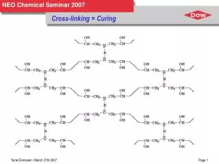
Corneal collagen cross-linking (CXL) • It is a rather new therapeutic approach attempting to address KC progression. • By using riboflavin (Vitamin B2) and ultraviolet A (UVA, 365nm) radiation. • The primary objective of CXL is to stabilize the collagen fiber matrix in KC corneas.
Corneal collagen cross-linking (CXL) • After surg-erery
Corneal melting • Postoperative corneal melts may be associated with infectious, inflammatory, or trophic causes. • The most common causes of corneal melt are herpes simplex virus (HSV) keratitis , retained lenticular materialand autoimmunedisorders.
Case presentation • 23-year-old Caucasian man. • He had progressive bilateral keratoconus. • He had no other ophthalmological problems. However, during the past year, he had developed contact lens intolerance. • UCVA : OD : 0.4 logMar OS : 0.5 logMar BCVA OD : 0.1 logMar (- 0.25 -2.50 X20)20/25 OS : 0.3 logMar (-0.50 – 3.0 X155)20/40
Central corneal pachymetry OD = 462μm OS = 455 μm • keratometric readings OD : K1= -43.1 , K2= -46.4 OS : K1= -43.2 , K2= -46.6 • The thickness of the thinnest corneal point (TCT) in the left eye was 443 μm
postoperative • during the first postoperative day the patient developed : • Intense photophobia. • Watering . • Non-specific ocular discomfort.
Slit lamp biomicroscopy • redness, especially at the limbal region, • severe corneal haze . • accompanied by non-specific endothelial • precipitates. • few inflammatory cells in the anterior • chamber. • patient’s visual acuity was limited to counting • fingers. • patient’s visual acuity was limited to counting fingers
Slit lamp biomicroscopic image showing severe corneal haze and endothelial precipitates due to the acute inflammatory response.
treatment • The patient's postoperative medication was modified to ofloxacin drops ,Exocin,Optive and Zovirax. • The treatment change resulted in subjective improvement of ocular discomfort and disappearance of the inflammatory cells in the anterior chamber. • However, the cornea presented extremely slow re- epithelialization and progressive thinning, which resulted in descemetocele and finally perforation in the second month.
Discussion • Corneal CXL isa temporary block in the progression of keratoconus . • the technique has an excellent safety profile if: • 1-De- epithelialization of the cornea to facilitate the absorption of riboflavin. • 2-Use of riboflavin 0.1% for at least 30 min. • 3-Homogeneous UV irradiation. • 4-A minimal central corneal thickness of 400 μm. • ( All of the criteria were met in our case )
corneal melting literature • An extensive literature search retrieved the following cases of CXL melting. • (Gokhale et al,2010) recently presented a case of acute corneal melting after CXL for keratoconus which was attributed to the hazardous impact of diclofenac on stromal keratocytes. Despite the fact that no apparent etiologic relationship between non-steroidal anti-inflammatory drugs (NSAIDs).
corneal melting literature • several investigators have attempted to associate keratolysis with postoperative NSAID therapy [Örnek K ,2008]. • The potential impact of NSAIDs on keratocytes is well known to the authors, thus we did not use NSAIDs as standard postoperative treatment in CXL. • bilateral melting after CXL for keratoconus in a patient with Down syndrome; however, the required minimal stromal thickness of 400 μm was not met.[Faschinger C ,2010]
in our case • Neither the central corneal thickness nor the thinnest corneal thickness was below 400 μm. • No evidence of non-infective keratitis. • No indications of hypersensitivity to riboflavin could be identified. • because of the shielding effect of riboflavin, the standard CXL procedure seems to cause no damage to the endothelial cells.
Conclusion • The exact cause of corneal melting in our case remains unknown to us. • An immunohistochemical examination of the affected cornea could provide more data regarding its pathological mechanism. • Nevertheless, since all precautions for standard CXL treatment were met in our case, • Further research is necessary to address all safety issues associated with this procedure.