Download

1 / 23
230 likes | 399 Views
Homeostasis and Endocrine Signaling. Tissues. Figure 32.2 4 major types: Epithelial – found on outside of the body and lining organs and cavities. Muscle – 3 types Cardiac – heart tissue, involuntary Smooth – involuntary actions in body, organs, blood vessels

E N D
Tissues • Figure 32.2 • 4 major types: • Epithelial – found on outside of the body and lining organs and cavities. • Muscle – 3 types • Cardiac – heart tissue, involuntary • Smooth – involuntary actions in body, organs, blood vessels • Skeletal – muscle that moves, attaches to bone, voluntary
Nervous tissue – the neuron, sends impulses, communication • Glia cells are nerve helping cells to the neurons • Connective tissue– diverse group of tissues scattered throughout body and extracellular matrix • Bone – calcified hard matrix • Blood – liquid matrix • Cartilage – ear, nose, gel like matrix • Dense fibrous – tendons and ligaments • Adipose - fat • Areolar – loose fibrous connecting tissue
Regulator or Conformer? • Animals that are regulators uses internal mechanisms to control internal change – endothermic, homeothermic, warm blooded • Animals that are conformers – internal condition changes in accordance with external changes, ectothermic, cold blooded • Homeostasis – maintenance of a constant internal balance • examples,- body temp, blood glucose levels… • Negative feedback – when body is out of homeostasis and it is brought back. • Positive feedback – when body is brought out of homeostasis purposely for a short period of time, childbirth and oxytocin
Thermoregulation (heat) • Figure 32.3
Figure 32.4 Sensor/ control center: Thermostat turns heater off. Response: Heating stops. Room temperature decreases. Stimulus: Room temperature increases. Set point: Room temperature at 20C Stimulus: Room temperature decreases. Room temperature increases. Response: Heating starts. Sensor/ control center: Thermostat turns heater on.
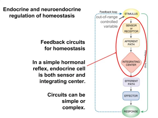
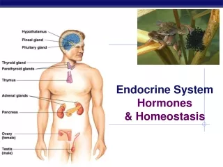
Endocrine system • Endocrine system – communication via hormones that are released by endocrine glands into the blood stream. • Hormones – chemical messengers • Exocrine glands – figure 32.11 • Exocrine glands – integumentary system, release product to cavity or outside the body, sweat. • Nervous system – rapid communication using neurons and nerve impulses • All run by Stimulus/Response mechanism
Figure 32.11a Major Endocrine Glands and Their Hormones Hypothalamus Pituitary gland Anterior pituitary Pineal gland Melatonin Posterior pituitary Oxytocin Vasopressin (antidiuretic hormone, ADH) Thyroid gland Thyroid hormone (T3 and T4) Calcitonin Adrenal glands (atop kidneys) Parathyroid glands Parathyroid hormone (PTH) Adrenal medulla Epinephrine and norepinephrine Ovaries (in females) Estrogens Progestins Adrenal cortex Glucocorticoids Mineralocorticoids Testes (in males) Androgens Pancreas Insulin Glucagon
Figure 32.8 Sensor/controlcenter: Thermostat in hypothalamus Response: Sweat Response: Blood vessels in skin dilate. Stimulus: Increased body temperature Body temperature decreases. Homeostasis: Internal body temperature of approximately 36–38C Body temperature increases. Stimulus: Decreased body temperature Response: Blood vessels in skin constrict. Sensor/controlcenter: Thermostat in hypothalamus Response: Shivering
Figure 32.9 (a) Signaling by hormones (b) Signaling by neurons Stimulus Stimulus Endocrine cell Cell body of neuron Nerve impulse Axon Hormone Signal travels to a specific location. Signal travels everywhere. Blood vessel Nerve impulse Axons Response Response
Osmoregulation (fluids) • How animals control solute concentrations in the interstitial fluid and balance water gain and loss • Excretory system – releasing of nitrogenous and metabolic waste products (kidney) • Osmoconformer – being isoosmotic with its surroundings, marine animals • Osmoregulator – to control internal osmolarity independent of the environment. Allows animals to live in freshwater/terrestrial habitats.
Nitrogenous wastes in animals • 32.16
Figure 32.16 Proteins Nucleic acids Amino acids Nitrogenous bases Amino groups Most aquatic animals, including most bony fishes Mammals, most amphibians, sharks, some bony fishes Many reptiles (including birds), insects, land snails Urea Uric acid Ammonia
Figure 32.16a Many reptiles (including birds), insects, land snails Most aquatic animals, including most bony fishes Mammals, most amphibians, sharks, some bony fishes Urea Ammonia Uric acid
The excretory process • Urine formation: 32.17
Figure 32.17 Filtration Capillary Filtrate Excretory tubule Reabsorption Secretion Urine Excretion
Figure 32.19b Kidney Structure Renal cortex Renal medulla Nephron Organization Renal artery Renal vein Afferent arteriole from renal artery Glomerulus Bowman’s capsule Ureter Proximal tubule Renal pelvis Peritubular capillaries Distal tubule Nephron Types Cortical nephron Efferent arteriole from glomerulus Branch of renal vein Descending limb Renal cortex Loop of Henle Vasa recta Collecting duct Ascending limb Renal medulla Juxtamedullary nephron
Figure 32.19bc Nephron Organization Afferent arteriole from renal artery Glomerulus Bowman’s capsule Proximal tubule Peritubular capillaries Distal tubule Efferent arteriole from glomerulus Branch of renal vein Descending limb Loop of Henle Vasa recta Collecting duct Ascending limb
Figure 32.20 4 5 3 2 1 3 Proximal tubule Distal tubule NaCI Nutrients H2O H2O HCO3− HCO3− K NaCI NH3 H H K Interstitial fluid CORTEX Thick segment of ascending limb Descending limb of loop of Henle Filtrate H2O NaCI Salts (NaCI and others) H2O HCO3− OUTER MEDULLA NaCI H Urea Thin segment of ascending limb Collecting duct Glucose, amino acids Some drugs Urea NaCI H2O Key INNER MEDULLA Active transport Passive transport
Adaptations • Based on where you live, there are adaptations to the kidney • Hyperosmotic urine (dessert animals) – long loops of Henle that extend deep into the medulla • Birds – shorter loop of Henle, les concentrated urine compared to mammals – uric acid is product to help conserve water.
Homeostatic regulation of kidney • 32.23 antidiruretic hormone
Figure 32.23-3 Osmoreceptors trigger release of ADH. Thirst Drinking of fluids ADH Increased permeability Distal tubule STIMULUS: Increase in blood osmolarity H2O reabsorption Collecting duct Homeostasis