The Parotid Gland: Anatomy and Function
200 likes | 361 Views
The comprehensive guide to the parotid region, covering the structure, subdivisions, relations, duct anatomy, nerve supply, blood supply, and lymphatic drainage. Explore the essential knowledge about the largest salivary gland in the body.

The Parotid Gland: Anatomy and Function
E N D
Presentation Transcript
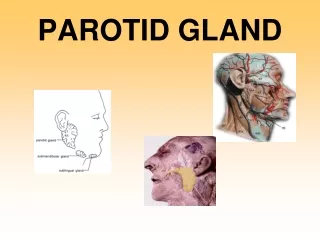
THE PAROTID REGION Dr. Ahmed Fathalla Ibrahim
THE PAROTID REGION • It includes: • The parotid salivary gland • The structures related to the gland
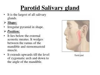
THE PAROTID GLAND • DEFINITION: It is the largest of the salivary glands • SITE: It lies below the auricle, occupying the region between ramus of mandible & mastoid process • EXTENT: • Superiorly: to zygomatic arch • Inferiorly: to angle of mandible • Anteriorly: to overlap posterior border of masseter • Posteriorly: to overlap anterior border of sternomastoid • SHAPE: Pyramidal
THE PAROTID GLAND • SUBDIVISIONS: • Main gland • Accessory gland: above parotid duct • CAPSULE: • Derived from deep fascia of neck (cervical fascia) • Its superficial layer is attached to zygomatic arch & extends to cover masseter • Its deep layer is attached to mandible, styloid & mastoid processes • A thickening of deep fascia extends from styloid process to angle of mandible (stylomandibular ligament) & separates the capsule of parotid from that of submandibular gland • It is tense (swellings of parotid gland are painful)
THE PAROTID GLAND • RELATIONS: • Superficial: skin, superficial fascia, great auricular nerve, superficial parotid (preauricular) lymph nodes • Anteromedial: posterior border of ramus of mandible + muscles attached to ramus (masseter, medial pteygoid) • Posteromedial:mastoid process + muscles attached to it (sternomastoid, posterior belly of digastric), styloid process + muscles attached to it (stylohyoid, styloglossus, stylopharyngeus), carotid sheath & its contents (internal jugular vein, internal carotid artery, 9th, 10th, 11th & 12th cranial nerves) • Medial: pharyngeal wall
STRUCTURES WITHIN THE PAROTID GLAND • Termination of facial nerve & beginning of its five terminal motor branches : most superficial structures • Terminations of superficial temporal & maxillary veins + the whole retromandibular vein + beginning of its two divisions (anterior & posterior) • Termination of external carotid artery & beginning of its two terminal branches (superficial temporal & maxillary): deepest structures • Deep parotid lymph nodes: embedded within substance of the gland
PAROTID DUCT • LENGTH: Two inches • COURSE & RELATIONS: • Emerges from anterior border of gland • Runs obliquely forwards, superficial to masseter & below transverse facial artery & accessory parotid • TERMINATION: • Pierces: buccal pad of fat, buccopharyngeal fascia, buccinator muscle & buccal mucosa • Opens: into the vestibule of mouth, opposite the crown of upper 2nd molar tooth • APPLIED ANATOMY: The oblique passage of the duct act as a valve-like mechanism & prevents inflation of the duct during blowing • SURFACE ANATOMY: It is represented by the middle 1/3 of a line extending from the tragus of the auricle to a point midway between the ala of nose & upper lip
NERVE SUPPLY • PARASYMPATHETIC (SECRETORY): • Origin: inferior salivary nucleus (medulla) • Preganglionic fibers: run along the lesser petrosal nerve (branch of tympanic of glossopharyngeal (9th cranial) • Ganglion: fibers relay in the otic ganglion (infratemporal fossa) • Postganglionic fibers: reach the parotid gland along auriculotemporal nerve (branch of mandibular of trigeminal) • SYMPATHETIC:Postganglionic sympathetic fibers reach the gland as a plexus around external carotid artery
BLOOD SUPPLY • ARTERIES: External carotid • VEINS: Retromandibular vein
LYMPHATIC DRAINAGE • Into superficial & deep parotid lymph nodes • Finally into deep cervical lymph nodes