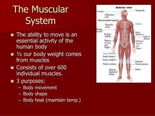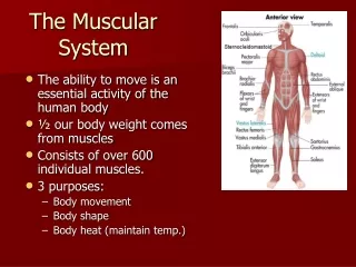
The Muscular System
560 likes | 582 Views
This article provides an overview of the skeletal, cardiac, and smooth muscles, including their structure, organization, and function. It also discusses muscle regeneration and specific muscles of the head.

The Muscular System
E N D
Presentation Transcript
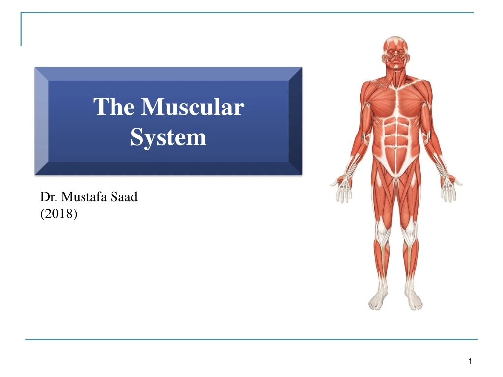
The Muscular System Dr. Mustafa Saad (2018)
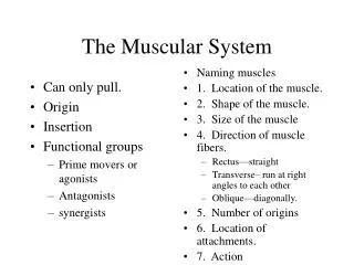
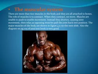
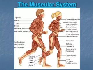
Skeletal muscles are formed of several bundles of skeletal muscle cells. They are attached by tendons to bones • When a skeletal muscle contracts, the tendon will be pulled and this will pull the bone resulting in Movement • The belly of the muscle is the fleshy (wide) part between the tendons • Muscles have more than one bony attachment: • the attachment of a tendon to the relatively stationary bone is called the origin. • the attachment of the muscle’s other tendon to the relatively movable bone is called the insertion. • the action/s of a muscle are the main movements that occur during contraction (e.g., flexion or extension).
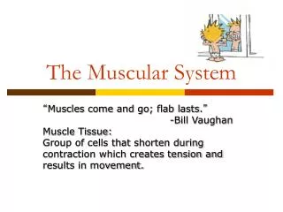
Muscular Tissue Muscular tissue is the type of tissue whose cells are differentiated to optimally use the contractile ability of the cells. Cell membrane = Sarcolemma Cytoplasm = Sarcoplasm Smooth endoplamsic reticulum = Sarcoplasmic reticulum
Types of Muscle Cells • Muscle cells are relatively long, therefore, they’re called muscle fibers • There are three types of muscle cells: Fig.1: Types of muscle cells.
Notes • Smooth muscle cells are held together by desmosomes. Also, gap junctions are present between the cells to allow the spread of Ca2+ (and thus contraction) rapidly between them. • The branches of cardiac muscle cells meat each other at specialized structures called the intercalated discs which also contain desmosomes and gap junctions. Fig.2: Cardiac muscle cells.
Organization of Skeletal muscles: • Skeletal muscles are formed of several bundles of muscle fibers. • Each fiber is surrounded by Endomysium: a loose areolar connective tissue layer. Each bundle is surrounded by connective tissue Perimysium. The whole muscle is surrounded by Epimysium: a dense connective tissue layer. • These three connective tissue layers will extend beyond the fleshy part of the muscle to form the cord-like tendons or the broad aponeuroses that attach muscles to bones.
Fig.3: Structure of skeletal muscles and their covering layers.
Cross-Striation of skeletal and cardiac muscle cells: • Skeletal and cardiac muscle fibers, under the LM, appear to have alternating dark and light areas. These are called the A and I bands respectively. The banding is due to the regular arrangement of the thin myofilamentActin and the thick myofilament Myosin. • Under the EM, this arrangement proves to be more complex. Fig.4: Striation under light microscope.
Fig.5*: Striation under electron microscope. • H Zone: a lighter colored area within the A band. • M Line: darker colored line in the middle of the H zone. • Z Disc (Line): a dark line in the middle of the light I band.
The Sarcomere: is the repetitive functional subunit of the contraction apparatus. It extends from one Z-line to the next Z-line. • Several sarcomeres arranged end-to-end form the cylindrical myofibrils. Each muscle fiber contain several myofibrils. Fig.6: Sarcomeres and myofibrils.
Muscle Regeneration • Skeletal muscle cells cannot divide. Inactive Satellite cells are present close to the muscle fibers. When injury occurs, the satellite cells become active, divide and form new skeletal muscle fibers. This is also thought to be the mechanism by which skeletal muscles hypertorphy after exercise. • Cardiac muscles lack satellite cells. After injury, the damaged muscles are replaced by a connective tissue scar. • Smooth muscle cells can divide, and, therefore, can easily replace damaged cells.
Muscles Of The Head Muscles Of Facial Expression • Muscles of facial expression: • Lie within the subcutaneous layer. • Usually originate from skull bones & insert into the skin. • Are all supplied by the Facial nerve. • Because of their insertions, the muscles of facial expression move the skin rather than a joint when they contract. Because of this, these muscle produce the wide variety of facial expressions that humans have.
Zygomaticus major – The muscle of true smile Risorius – The muscle of false smile (probably present only in humans and gorillas) Fig.7*: Muscles of facial expression.
Muscles of Mastication (Chewing) • Four pairs of muscles move the mandible, and are known as ‘muscles of mastication’. • They are all supplied by the mandibular branch of the trigeminal nerve. • The masseter, temporalis, and medial pterygoid close the mouth and account for the strength of the bite. • The medial and lateral pterygoid muscles help to chew by moving the mandible from side to side. • The lateral pterygoid is also the main depresseser of the mandible as in opening the mouth. Note that Gravity assists in depressing the mandible (plus other muscles).
Muscles Of The Tongue • Muscle of the tongue include: • Intrinsic muscles (originate and insert within tongue). These are responsible for changing the shape of the tongue. • Extrinsic muscles (originate outside the tongue, insert into tongue). These are responsible for moving the tongue. • Genioglossus is one of these extrinsic muscles. It moves the tongue forwards. • All muscles of the tongue are supplied by the Hypoglossal nerve, except the palatoglossus. Fig.9: Muscles of the tongue.
Muscles Of The Neck • TheSternocleidomastoid (SCM) muscle is an important anatomical landmark in the neck. It divides the neck into an anterior and a posterior triangle. • The SCM muscle arises from the sternum and clavicle and is inserted into the mastoid process and the occipital bone. Its motor supply is by the accessory (XI) nerve. If the muscles on both sides contract, they’ll flex the head. If the SCM muscle of one side contracts, it’ll rotate the head to the opposite side.
Anterior Triangle: • Anterior border: midline • Posterior border: SCM muscle • Superior border: Mandible • Posterior Triangle: • Anterior border: SCM muscle • Posterior border: Trapezius muscle • Inferior border: Clavicle Fig.11: Boundaries of the triangles of the neck.
Muscles of the anterior part of neck: • Two main muscle groups:
Lateral Vertebral Muscles – The Scaleni: • These are attached to the cervical part of the vertebral column and pass laterally • Scalenus anterior is an important landmark in the neck with several important relations. Among these relations we have: the subclavian artery and vein and the trunks of the brachial plexus
Respiratory Muscles Of The Thorax • Respiratory muscles alter the size of the thoracic cavity which affects the pressure in the lungs, and that determines whether we inhale or exhale. • Between the ribs we have the Intercostal muscles arranged in three layers: the External, Internal and Innermost intercostal muscles. Between the internal and innermost intercosal muscles, we have the intercostal nerve and vessels. • There are also a number of accessory muscles useful in forced breathing: SCM and the scaleni muscles.
The Diaphragm The diaphragm is the most important muscle of respiration
Anterior Abdominal Wall Muscles • The anterolateral abdominal wall muscles include the external oblique, internal oblique, and transversusabdominis muscles which form three protective layers around the abdomen. • The muscle bundles of each layer extend in a different direction, offering great protection to the abdominal viscera. • The aponeurosis (broad tendon) of the external oblique forms the thick inguinal ligament inferiorly. • The aponeuroses of these 3 muscles form the rectus sheaths which enclose the rectus abdominis muscles. • The sheaths meat each other in the midline to form the linea alba, a connective tissue band extending from the xiphoid process to the pubic symphysis.
Actions: • They retain the organs within the abdominal cavity. • The oblique muscles laterally flex and rotate the trunk. • The rectus abdominis flexes the lumbar vertebrae. • By contracting simultaneously with the diaphragm, they increase intra-abdominal pressure and help in micturition, defecation, vomiting, and labor. • They may contract at the end of expiration, pushing the relaxed diaphragm further upwards into the thorax.
Posterior Abdominal Wall Muscles Quadratus lumborum: depresses 12th rib Flex thigh on trunk. If thigh is fixed, flex trunk on thigh Fig.17*: Posterior abdominal wall muscles.
Muscles of the Back • Muscles that move the backbone are quite complex having multiple origins/insertions with considerable overlap between the muscles. • They include several groups of muscles whose main function is the (1)extension of the spine and (2)the maintaining of posture. Fig.18: Deep muscles of the back.
Muscles Of The Upper Limb Muscles that move the Pectoral Girdle • Subclavius: attached to clavicle • Levator scapulae, Rhomboid major and rhomboid minor Trapezius: Is a large muscle seen on the back. With the serratus anterior muscle, it rotates the scapula so that its glenoid cavity is raised. This allows the arm to be raised above the head. Serratus anterior (Punching muscle): fix scapula in position Pectoralis minor • These muscles also stabilize the girdle so that the free limb can have a firm base to move on
Fig.19: Muscles working on the pectoral girdle – anterior view.
Fig.19: Muscles working on the pectoral girdle – posterior view.
Rotator Cuff Muscles Fig.21: Rotator cuff muscles. • The tendons of these muscles all blend with the capsule of the shoulder joint, thus help in stabilizing it. • The supraspinatous helps initiate abduction of the arm.
Muscles of the arm (that move the forearm) • The biceps brachii, brachialis, and brachioradialis are flexors. The triceps brachiiextends the forearm • The biceps has two heads of origin. The long head passes through the intertubercularsulcus of the humerus. The biceps inserts into the radial tuberosity. It also forms an aponeurosis that inserts medially into fascia and that protects the underlying brachial artery and median nerve as they pass in the cubital fossa.
Fig.23: The cubital fossa. The Cubital Fossa: • Shallow triangular depression anterior to elbow joint. • Tendon of biceps, brachial artery and median nerve pass through it. • Site of measuring brachial artery pulse and taking blood pressure. • The superficial veins passing in the skin overlying this fossa can be used to take blood samples.
Muscles of the Forearm • Muscles in this group that act on the wrist and fingers are known as extrinsic muscles of the hand because they originate outside the hand and insert within it. • Based on location and function, these muscles are divided into an anterior (flexor) compartment and a posterior (extensor) compartment. • Anconeus, supinator and pronator quadratus are muscles in the forearm that act on the forearm.
Fig.24*: Muscles of the anterior compartment of the forearm.
As the long muscles of the anterior forearm pass over the carpal bones, they are held in place by a thick band of connective tissue called the flexor retinaculum (transverse carpal ligament). This band with the carpal bones form a tunnel called the carpal tunnel. • Also passing through this tunnel is the median nerve. • Certain conditions may affect this tunnel (like inflammation of the tendons or the joints) leading to compression of the median nerve. This is called Carpal Tunnel Syndrome. Fig.25: The carpal tunnel. • The affected person may have pain in the hand, change in sensations and even weakness in the hand muscles supplied by the median nerve.
Intrinsic Muscles of the Hand (3 groups) • The intermediate group include the lumbricals, the palmar and dorsal interossei. • The palmar interossei adduct the fingers towards the middle finger. The dorsal interossei abduct the fingers away from the middle finger Hypothenar muscles act on the little finger Thenarmuscles act on the thumb Fig.26*: Intrinsic muscles of the hand.
Muscles Of The Lower Limb • Lower limb muscles function in stability, locomotion, and maintenance of posture. In contrast, upper limb muscles are characterized by versatility of movement. • Muscles of the lower limbs often cross two joints and can act equally on both. • Most muscles that move the femur originate from the pelvic girdle and insert on the femur.