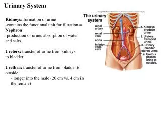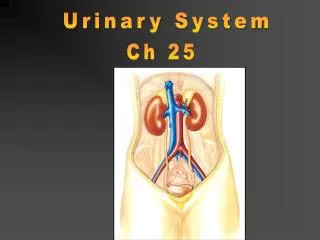
URINARY SYSTEM
URINARY SYSTEM. OVERVIEW. Urinalysis Serum/plasma urea & creatinine concentrations Urine protein to creatinine ratio (UPC ratio). 1.URINALYSIS. MAIN INDICATIONS ARE: • Evaluation of renal & lower urinary tract abnormalities • Assessment of some metabolic/endocrine disorders


URINARY SYSTEM
E N D
Presentation Transcript
OVERVIEW • Urinalysis • Serum/plasma urea & creatinine concentrations • Urine protein to creatinine ratio (UPC ratio)
1.URINALYSIS • MAIN INDICATIONS ARE: • • Evaluation of renal & lower urinary tract abnormalities • • Assessment of some metabolic/endocrine disorders • •Assessment of state of hydration
Gross evaluation Specific gravity (SG) Urinalysis Biochemical analysis Sediment examination (microscopy) Culture
GROSS EVALUATION: COLOUR Urine from healthy animals can vary in colour but is usually light, mild or dark yellow Red discolouration Red discolouration may indicate haemoglobinuria, myoglobinuria or haematuria.
GROSS EVALUATION: TURBIDITY turbidity in urine from a cow with pyelonephritis Upper sample :Clear urine Lower sample:Turbid Turbid urine (healthy horse)
TURBIDITY • Healthy horses and rabbits may have turbid urine due to high concentration of mucin and crystals. In other species turbidity can indicate the presence of sediment. • On refrigeration, urine samples may become turbid from crystallisation of minerals which were in solution, and they may clear when returned to room temperature
Gross evaluation Specific gravity (SG) Urinalysis Biochemical analysis Sediment examination (microscopy) Culture
SPECIFIC GRAVITY (SG) Ratio of weight (density) of urine to that of an equal volume of water at the same temperature. No units. Values depend on: - hydration status and water intake - the kidney’s concentrating ability It is a test of renal tubular function Hydration status can be determined by assessing skin turgor or by measurement of serum albumin, or PCV and total proteins.
SPECIFIC GRAVITY (SG): MEASUREMENT • Reagent test strips are unreliable for animals/Always use the refractometer • If the urine is turbid, centrifuge it before measuring SG of the supernatant 1.050 1.040 1.030 1.020 1.010 1.000
SG: INTERPRETATION • HYPERSTHENURIA: concentrated urine • - >1.012 • -urine of healthy, normally hydrated animals • ISOSTHENURIA: urine neither concentrated nor diluted • -1.007-1.012 (urine SG = plasma filtrate SG) • -persistent isosthenuria warrants further investigation • HYPOSTHENURIA: urine is more diluted than plasma • - <1.007 • - persistent hyposthenuria warrants further investigation
SG: INTERPRETATION • The range of values for SG can vary according to water intake and hydration status. Usually SG in normal concentrated urine is >1.030 • WATER DEPRIVATION TEST • Contra-indication: It should never be carried out in depressed, dehydrated or azotemic animals, or if renal failure is suspected. • Indication: Confirmation of the animals ability to concentrate its urine when water is withheld. • Protocol:The urine SG is monitored every 2 hours until 5% of body weight is lost, or the urine SG is >1.020. • Interpretation: • If the urine SG increases to 1.020, tubular function and ADH availability are confirmed. • If the urine SG remains <1.020, diabetes insipidus is suspected.
Gross evaluation Specific gravity (SG) Urinalysis Biochemical analysis Sediment examination (microscopy) Culture
BIOCHEMICAL ANALYSIS URINE STRIPS Always follow manufacturer instructions
- Glucose is not normally found in urine of healthy animals - Causes of glycosuria • Persistent hyperglycaemia - Diabetes mellitus • Transient hyperglycaemia - Stress in cats - Drugs (xylazine, ketamine) - IV fluids containing glucose - Convulsions • Renal tubular disorders - Fanconi syndrome - Primary glucosuria GLUCOSE
BILIRUBIN • Not accurate for dogs/cats • Tests utilising a tablet (ictotest) can be more accurate than strip-tests • Light can break down bilirubin • Trace to + normal in healthy dogs.No bilirubin present in the urine of other healthy animals • The bilirubin in the urine is water-soluble conjugated bilirubin • Causes of bilirubinuria • Same as causes of bilirubinaemia
KETONES • Accurate test for animals • Does not detect -hydroxybutyric acid • Ketones are not present in the urine of clinically healthy animals •Trace can be normal in rabbits • Causes of ketonuria •Diabetes mellitus, pregnancy, starvation, ketosis, immediately after calving in high–producing dairy cows N.B. Many disorders causing anorexia in cattle (e.g. mastitis, metritis, pneumonia) will cause ketonaemia and ketonuria, but levels of ketones are generally not as high as in primary ketosis.
BLOOD / HAEMOGLOBIN - Accurate test for animals - Detects intact RBCs, haemoglobin or myoglobin • Follow-up positive result with sediment examination • Interpret positive result in conjunction with the method of urine collection (cystocentesis can be a cause of presence of blood in urine)
pH • Acceptable test for animals • Carnivores: • acidic urine is normal if fed a meat diet • alkaline urine usually reflects urinary tract infection - Herbivores: • alkaline urine is normal • acidic urine may reflect increased protein catabolism e.g. high protein diet, starvation, fever, nursing animals • Some drugs can influence pH • Not an accurate indicator of systemic acid/base balance
PROTEIN • Acceptable test for animals but can give false positive reaction in alkaline samples. • Test detects mainly albumin. Does not detect globulins • Always interpret in conjunction with SG and sediment examination (it is not abnormal to have trace protein in concentrated urine but always abnormal finding in diluted urine). • Common causes of proteinuria: • urogenital haemorrhage • urogenital inflammation • renal protein loss
NITRITE, UROBILINOGEN, LEUKOCYTES - Nitrite • Positive results may indicate bacterial infection • false negative results occur commonly - Urobilinogen • Questionable clinical usefulness - Leukocytes • False negative results common in dogs • False positive results common in cats NONE ARE RELIABLE IN EXAMINING ANIMAL URINE
UREA LIVER BLOOD Urea cycle Urea TISSUES Urea NH4+ 75% NH4+ KIDNEYS proteins 25% Urea Urea Dietary Proteins bacteria NH4+ Urea in urine GASTROINTESTINAL TRACT
UREA and creatinine • Glomeruli: 75% of urea is excreted (excretion or when glomerular filtration rate or ) • Tubules: Urea is reabsorbed (reabsorption or when glomerular filtration rate or ) • Creatinine is derived from creatine-phosphate, creatinine is excreted via the glomeruli. It is not reabsorbed in the tubules so excretion of creatinine is a measure of glomerular filtration rate.
BUN vs. UREA • BUN = blood urea nitrogen= concentration of the nitrogen component of urea in blood • BUN value is Lower than urea value. BUN:Urea ratio is approximately 1:2 • But the term BUN is used interchangeably with urea
CREATININE KIDNEYS creatinine creatinine creatine MUSCLE creatinine BLOOD Creatinine in urine NH4+ creatinine INTESTINES
AZOTAEMIA - Increased serum/plasma urea & creatinine concentrations URAEMIA • Marked azotaemia and clinical signs (vomiting, anorexia, gastrointestinal ulceration)
AZOTAEMIA • CAUSES • PRE-RENAL • RENAL • POST-RENAL
PRE-RENAL AZOTAEMIA - DECREASED RENAL PERFUSION - Hypovolaemia, dehydration, cardiovascular disease • Urea is and creatinine Normal / • Urine specific gravity is - INCREASED UREA PRODUCTION - G.I. TRACT HAEMORRHAGE • Urea is and creatinine is Normal - HIGH PROTEIN DIET • Urea is and creatinine is Normal
RENAL AZOTAEMIA - RENAL DISEASE - ONLY evident when more than 60-75% of nephrons are compromised • Urea and creatinine are • Urine is inadequately concentrated
POST-RENAL AZOTAEMIA - URINARY TRACT OBSTRUCTION • ureter, urethra - URINARY TRACT RUPTURE • ureter, bladder, urethra - Urea and creatinine are
LOW SERUM/PLASMA UREA: CAUSES • Decreased Liver Function • Portosystemic shunt • Increased Excretion • Extreme PU/PD • Overhydration • Low protein intake Young Animals have a lower reference range
RUMINANTS / HORSES - Excrete most of urea via the gut (very little via kidneys). So blood urea can be normal despite severe renal disease. - Therefore blood creatinine is a more sensitive indicator of renal disease
UPC RATIO • Used to assess the clinical significance of proteinuria • Total protein & creatinine concentrations are measured in a single urine sample and expressed in the same units