Urinary System
Urinary System. Urinary System Structure Urine Formation Regulating Urine Formation. 10.1 Urinary System Structure. Kidneys Nephrons Capillary Beds. Kidneys. The kidneys lie in the lower, dorsal part of the abdominal cavity


Urinary System
E N D
Presentation Transcript
Urinary System Urinary System Structure Urine Formation Regulating Urine Formation
10.1 Urinary System Structure Kidneys Nephrons Capillary Beds
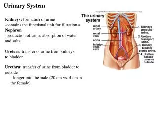
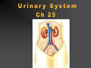
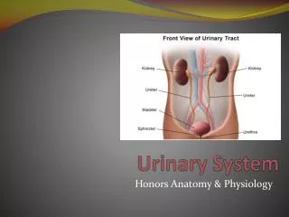
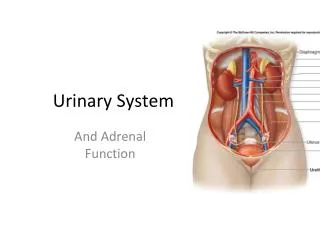
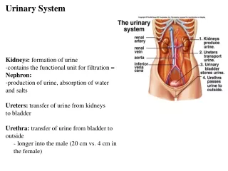
Kidneys • The kidneys lie in the lower, dorsal part of the abdominal cavity • They receive blood from the renal arteries, remove the wastes from it, and return the “clean blood” to the circulatory system • The actual waste removal from the blood and production of urine are functions of microscopic structures called nephrons(each kidney contains millions)
Nephrons • Urine collects in the pelvic region of each kidney before peristalsis conducts it to the urinary bladder • When the urinary bladder is filled, the urination response is triggered, and the fluid is excreted • Nephrons are the functional units of the urinary system; these specialized tubules originate in the cortex of the kidneys
The structure of every nephron follows the same pattern, each one beginning with a “blind end” called Bowman’s capsule • From there they twist back and forth before they loop down into the renal medulla • Each tubule loops back up to form a second twisted region before joining a common collecting duct
The three regions of each nephron between Bowman’s capsule and the collecting duct are (in sequence): proximal convoluted tubule, loop of henle, distal convoluted tubule
Capillary beds • Blood (carrying wastes) is conducted into the cortical region of the kidney by branches of the renal artery called afferent arterioles • The capillary beds associated with the nephrons are highly specialized into two parts
Glomerulus • A tuft of extremely delicate capillaries that are enveloped by Bowman’s capsule • From the glomerulus, an efferent (outgoing) arteriole conducts blood to the second part of the capillary bed
Peritubular capillaries • Extend around the rest of the nephron • Eventually, the capillaries join to form a venule, which conduct blood back into the renal vein
10.2 Urine Formation Parts of the Nephron Video
Bowman’s Capsule • Urine formation is the process of a greatly modified version of capillary fluid exchange • Blood entering the cortex of the kidney is slowed down as it courses through the tiny delicate capillaries of a glomerulus
The glomerulus’ structure allows blood pressure to force small components of plasma of the blood into the extracellular fluid in the spaces surrounding the glomerulus • This space is occupied by Bowman’s capsules, which cup each glomerulus • Substances are forced into this porous section of the nephron by constant blood pressure
This process is called pressurefiltration; the fluid that enters the nephron tubules is called the filtrate • Blood cells and large molecules in plasma like globulins do not normally get forced into the filtrate because of their size, which helps to establish an osmotic gradient between the blood and the extracellular fluid
Proximal Convoluted Tubule • The filtrate contains some materials that are still useful and required by the body. • The cells that line the proximal convoluted tubule are specially designed with carrier proteins to pump theses needed materials back into the peritubular capillaries
Thus molecules like glucose, amino acids and other nutrients are returned to the blood • This activity, called selective reabsorption, requires ATP (the removal of substances by selective reabsorption makes the filtrate less concentrated) • Na+ is pumped (ATP) out of the filtrate, while Cl- and water (small amount) follow passively and are returned to the more concentrated plasma
Descending Loop of Henle • The nephron with its filtrate makes three passes between the cortex and the medulla before the urine enters the renal pelvis • During the first pass, the filtrate goes from the proximal convoluted tubule to the medulla through the descending loop of Henle • Cells at this portion of the loop are permeable to H2O, but not Na+
Because the loop encounters an extracellular environment with a high solute concentration as it descends into the medullary region, increasing amounts of H2O are removed • This H2O moves into the blood due to the osmotic gradient; filtrate is very “salty”
Ascending Loop of Henle • The ascending portion is permeable to Na+, but not to H2O • Because of the high Na+ of the filtrate concentration relative to the extracellular fluid, Na+ diffuses out of the filtrate • This feature contributes to the osmotic gradient that moves water out of the descending side
As the filtrate moves towards the cortex, the potential for diffusion lessens as the sodium ion concentration gradient lessens; ATP is used to remove additional Na+ from the filtrate • By the time the filtrate gets to the distal convoluted tubule in the cortical region of the kidney, it has been diluted by the removal of lots of Na+ making it iso-osmotic to the extracellular fluids
Distal Convoluted Tubule • During the process of tubular secretion, substances that are in excess in blood (penicillin, histamines, vitamins, etc.) can be pumped out of the peritubular capillaries around the distal convoluted tubules into the filtrate • It is in this manner that final adjustments to the composition of the blood are made • The pH of the blood in the distal tubule is adjusted by the differential reabsorption of H+ and HCO3-
Collecting Duct • In the final phase of urine formation, the filtrate is once again subjected to the increasing concentration gradient of the extracellular fluids as the collecting duct passes through the medulla • Because the filtrate was diluted by the removal of Na+, the extracellular fluid in the medulla is hypertonic to the filtrate and additional water is withdrawn as the filtrate moves to the renal pelvis • Normally about 99% of the water molecules in the filtrate are reabsorbed during urine production
Review • Tubular secretion of histamines occurs in the • loop of Henle • collecting duct • distal convoluted tubule • proximal convoluted tubule.
Tubular secretion of histamines occurs in the • loop of Henle • collecting duct • distal convoluted tubule • proximal convoluted tubule.
In which kidney region are the majority of Bowman's capsules found? • Renal vein • Renal pelvis • Renal cortex • Renal medulla
In which kidney region are the majority of Bowman's capsules found? • Renal vein • Renal pelvis • Renal cortex • Renal medulla
In which blood vessel of the kidney can the highest concentration of metabolic wastes found? • Renal vein • Renal artery • Efferent arteriole • Peritubular capillary bed
In which blood vessel of the kidney can the highest concentration of metabolic wastes found? • Renal vein • Renal artery • Efferent arteriole • Peritubular capillary bed
The pH of blood is increased by the movement of hydrogen ions from the • distal convoluted tubule into the peritubular capillaries • peritubular capillaries into the distal convoluted tubule • peritubular capillaries into the proximal convoluted tubule • proximal convoluted tubule into the peritubular capillaries
The pH of blood is increased by the movement of hydrogen ions from the • distal convoluted tubule into the peritubular capillaries • peritubular capillaries into the distal convoluted tubule • peritubular capillaries into the proximal convoluted tubule • proximal convoluted tubule into the peritubular capillaries
The cells that line the proximal convoluted tubule are specialized for • Filtration • Transport • Secretion • contraction
The cells that line the proximal convoluted tubule are specialized for • Filtration • Transport • Secretion • contraction
10.3 Urine Formation Urea Glucose
Urea • Throughout the process of urine formation, urea is minimally reabsorbed into blood because of its low threshold level in plasma • Some urea diffuses out of the collecting duct in the medullary region contributing to the high solute concentration of the fluid surrounding the loop • In this manner, urea contributes to the osmosis of water into the spaces on the descending side
Water is reabsorbed by blood and is recovered • Urea leaks back into the ascending side of the loop; the product remaining in the nephron tubule is urine • Urine is still mostly water even though by this time most of the water that entered the nephron has been reabsorbed
Glucose • Has a higher threshold level in plasma, therefore, after it enters the nephron, it is readily transported back into blood • The reabsorption of glucose requires the presence of ATP and proteins along the walls of the tubules (there are many) • Normally all the glucose can be reabsorbed into the blood because the liver removes any excess and stores it as glycogen, maintaining its low plasma concentration
Abnormally large and frequent urine flows are symptomatic of diabetes due to more sugar being in urine and hence less water reabsorbed by the blood • A diuretic is something that increases urine flows; for diabetics, insulin treatments are an antidiuretic
The requirements of the body and the conditions under which the body works are not constant; therefore the functioning of the urinary system cannot be constant
ADH • Antidiuretic hormone (ADH) is secreted by the posterior pituitary gland in response to diminished blood volume • This hormone occupies receptor sites on the collecting ducts, which increase its permeability to water; therefore more water us reabsorbed into the blood • In this manner, ADH counteracts low blood volume by decreasing the volume of urine production
Diuretics • Alcohol is a diuretic because it inhibits ADH production thus promoting larger urine volumes • Coffee is also a diuretic because it increases the blood volume by causing water retention; this increases pressure filtration and therefore promotes larger urine flows • Excessive consumption of diuretics can lead to dehydration
Other Conditions • Should blood pressure drop, perhaps due to low Na+ concentration resulting in less water in plasma, the whole process may be in jeopardy • Conditions like this set off a chain reaction ending with the release of the hormone aldosterone from the adrenal cortex • Aldosterone increases the amount of Na+reabsorption at the distal convoluted tubule and thus increases the retention of Na+ in plasm ( K+ excreted)
The effect of this is the same as that of ADH – more water is reabsorbed and urine becomes more concentrated • This compensates for the initial low blood pressure
Review • Which part of a nephron is MOST affected by ADH? • Loop of Henle • Collecting duct • Distal convoluted tubule • Proximal convoluted tubule.
Which part of a nephron is MOST affected by ADH? • Loop of Henle • Collecting duct • Distal convoluted tubule • Proximal convoluted tubule.
High solute concentration in the blood (hypertonic blood) is detected by the • Glomerulus • distal tubule • Hypothalamus • posterior pituitary gland.
High solute concentration in the blood (hypertonic blood) is detected by the • Glomerulus • distal tubule • Hypothalamus • posterior pituitary gland.
Which of the following most correctly describes the site of urea entry into blood and site of urea removal from blood? • Entry at liver, removal at liver • Entry at kidneys, removal at liver • Entry at liver, removal at kidneys • Entry at muscles, removal at kidneys.
Which of the following most correctly describes the site of urea entry into blood and site of urea removal from blood? • Entry at liver, removal at liver • Entry at kidneys, removal at liver • Entry at liver, removal at kidneys • Entry at muscles, removal at kidneys.