Cell modelling using the Atomic Force Microscope Gary Johnston
180 likes | 333 Views
Cell modelling using the Atomic Force Microscope Gary Johnston. Presentation Overview. Who and what – A brief Background Why – Are we doing this? How – are we planning on doing the work? Results – Some recent tentative results. Current Issues - near and not so near. Brief Background.

Cell modelling using the Atomic Force Microscope Gary Johnston
E N D
Presentation Transcript
Cell modelling using the Atomic Force Microscope Gary Johnston
Presentation Overview • Who and what – A brief Background • Why – Are we doing this? • How – are we planning on doing the work? • Results – Some recent tentative results. • Current Issues - near and not so near.
Brief Background • 2 BSc. Degrees, 1 in Molecular biology and 1 in computing. • A foot in both camps • Predominately a Programming background • Games background PS1, PS2 and PC. • Limited timeframes, limited processing power. • Moved to flight simulation around 2000 – Worked on various projects including Eurofighter and GR4 Tornado.
What – am I doing? • Trying to make sense of the data the biologists provide and build a useful statistical model from it. • The data comes from 2 sources. • The Atomic Force Microscope and the Confocal microscope. Actin stained PNT2 cells in a monolayer AFM Force curve Force vs Indentation
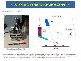
Atomic Force Microscope(AFM) • The AFM in a nutshell • We poke the cells with the stick and measure the force from the cantilever.
Atomic Force Microscope(AFM) • The AFM in a nutshell • The scale we are looking at is around a nanometer. • Very small!
Confocal microscope Confocal Actin stained PNT2 cell from a monolayer • Density Actin • Morphology of the Cell • Relative differences of irradiated Actin.
Why – Are we doing this? Radiation Study. • Study the effects of radiation on cancerous and non-cancerous cells. • In particular the how the cell cytoskeleton changes with dosage. • Rationale -: Since the actin cytoskeleton is thought to play a primary role with force within the cell, any changes should alter the force response for the cell • The two techniques mentioned earlier are used. • Confocal imaging – Distribution, morphology. • AFM - Force response.
How – are we planning on doing the work? The ‘Original’ Hertz Model (Conical) • Collecting all the constants together. • Becomes • Assumptions -: Linear varying E • So if we let a0 = 1... • if d = 2 , force = 4 N
How – are we planning on doing the work? • How do we know it’s not correct. • Below shows the best fit for a hertz curve verses that for a cell. N Force m Distance
How – are we planning on doing the work? • If we presume that the youngs modulus E varies non-linearly. • We come up with an extended hertz model. • Below is the fit for both the original and the extended model. Non-linear extended model. original
How – are we planning on doing the work? • The original hertz model was not acceptable. • The extended hertz model, gives a way of recording the force from a cell with reasonable accuracy over the span for the curve. • The next stage of this work is to use Ansys or MatLab. • To enable me to make a non-linear finite element model. • I’m hoping progress it to a data driven, dynamic model eventually. • Confocal data - used to influence the distribution and overall shape. • Force data - used to model the overall force response of the cell.
Recent results Tentative Initial result of investigation into effect of confluence. N • Result 1 – PC3 Confluent cells are less stiff • Test performed via Mann-Whitney equivalent in Matlab. • 95% likelihood that populations are not from the same sample. IE 95% Likely that they are statistically different. • This result requires more data to confirm that this is indeed a pattern for these particular cells. • To the right the Blue X represents the DAY 1 PC3 cells which are not confluent, whereas the red circles represent the DAY 2 PC3 cells which were confluent. Force/N
Time as a factor for AFM stiffness. • Result 2 – Time appears has no influence the cell stiffness for at least an hour. • The line of best fit has been plotted here and is roughly a straight line, this is based upon the force curves at 3x10⁻⁶ m indentation over 60 minutes in this case. • As can be seen the amount of force required doesn’t vary significantly. paras = (2*tan(37.5) ) / (0.75 * pi); Eb = p(3)/paras; k2 = p(2)/paras; k1 = p(1)/paras;
Time as a factor for AFM stiffness. • Result 2 – Eb, K1, K2 appear not to vary with time. This is in line with the result of the previous slide. • Shown here are the variations of EB,K1,K2 over the same interval. • As can be seen the least square line is only one of a number of fits you could do. • There appears to be no difference for cell stiffness over time, which is good as otherwise we’d have to factor time into the AFM results.
Summary • Build statistical model • From AFM and Confocal 3D images. • Look for statistical differences in response to radiation • between non-cancerous and cancerous cells. Current Issues (last as I’m Irish) • In the short term. • Choosing the right tool, Ansys or MatLab for modelling work. At present we are probably going to go for Ansys.
Current Issues (Longer Term) • Comparing populations of cells by Eb,K1,K2. Very complex. • Is it better to compare force populations at specific indentations and interpolate between fixed points?
Finally putting it all together. + Biology Where making random mistakes is common practice. = = The End !