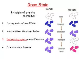
Download
1 / 5
50 likes | 60 Views
Acridine orange staining is a methodology for nucleic acid selective<br>metachromatic staining that is useful for following cell cycle determination. The interaction of acridine orange with target molecules (i.e. DNA<br>and RNA) is by intercalation or electrostatic attraction, respectively. This<br>work presents methodology and results of a cell staining that utilizes acri- dine orange fluoresence. It is possible to visualize under dark field ultra- violet microscope the actual viral infected host cells. The color production is stark and easily selected. This method appears to be versatile for a<br>number of various cell types in tissue culture, including human lympho- cytes. This methodology is demonstrated for Epstein Barr Virus (EBV), human immunodeficiency virus (HIV), and laboratory strain of lymphad- enopathy virus (LAV). The results of staining are reproducible and useful<br>for following viral infection and proliferation in tissue culture.

E N D
id1725875 pdfMachine by Broadgun Software - a great PDF writer! - a great PDF creator! - http://www.pdfmachine.com http://www.broadgun.com Volume 16 Issue 3 ISSN : 0974-7419 Analytical Analytical CHEMISTRY Analytical Analytical CHEMISTRY An Indian Journal Full Paper Full Paper ACAIJ, 16(3) 2016 [114-118] Acridine orange staining of virus infected host cells to monitor prolif- eration of viralinfection Ronald Bartzatt University of Nebraska, Durham Science Center, 6001 Dodge Street, Omaha Nebraska 68182, (USA) Tissue culture work and staining completed at the University of California-San Diego, San Diego, California, (USA) E-mail: rbartzatt@unomaha.edu ABSTRACT KEYWORDS Acridine orange stain; Virus; HIV; Infection; LAV. Acridine orange staining is a methodology for nucleic acid selective metachromatic staining that is useful for following cell cycle determina- tion. The interaction of acridine orange with target molecules (i.e. DNA and RNA) is by intercalation or electrostatic attraction, respectively. This work presents methodology and results of a cell staining that utilizes acri- dine orange fluoresence. It is possible to visualize under dark field ultra- violet microscope the actual viral infected host cells. The color produc- tion is stark and easily selected. This method appears to be versatile for a number of various cell types in tissue culture, including human lympho- cytes. This methodology is demonstrated for Epstein Barr Virus (EBV), human immunodeficiency virus (HIV), and laboratory strain of lymphad- enopathy virus (LAV). The results of staining are reproducible and useful for following viral infection and proliferation in tissue culture. 2016 Trade Science Inc. - INDIA INTRODUCTION counting[5], and observation and identification of nucleic acids[6]. AO has been applied to studies of vaginal and cervical cancer tissue[7], the study of blood parasites[8], and instrumental automated tech- niques[9]. AO is used effectively to examine wet mounts for diagnosis of T. vaginalis infection[10]. Clinicians have used AO to correctly diagnose for T. vaginalis for a multitude of patients. The staining methodol- ogy provides a manner for early detection of T. vaginalis[10]. AO staining does make it possible to distinguish acidic vesicles with different internal pH but is phototoxic which may lead to bursting of dye loaded vesicles[11]. Acridine Orange (AO) interacts with DNA and RNA by intercalation and electrostatic attraction, respectively[1]. AO is cell permeable and can inter- act with nucleic acid. DNA double strand intercala- tion fluoresces green while RNA electrostatic bound single strand fluoresces red[1]. Acridine orange stain- ing may also be useful for measuring apoptosis[1]. When illuminated by ultraviolet light and under dark field microscopy, the viral particles, whether single strand or double strand, will be illuminated. AO is the most popular of fluorochromes uti- lized for studies on whole blood[2, 3, 4], reticulocyte
ACAIJ, 16(3) 2016 Ronald Bartzatt et al. 115 Full Paper Full Paper AO staining has been applied to the rapid screen- ing of urine for significant bacterial presence in the automated TMS system, with success[12]. AO stain- ing methods have been used parallel gram staining to successfully describe and characterize infectious keratitis[13]. AO staining has been used successfully for early detection of positive blood cultures[14]. Staining using AO has been shown to be superior to gram stain for detection of microoganisms in cere- brospinal fluid and various clinical specimans[15]. AO staining has also been successful in the clinical detection and diagnoses of Trichomonas vaginalis[16]. The method is utilized successfully for vaginal discharge examination for T. vaginalis[17]. AO staining has been used to successfully detect the presence of bovine respiratory syncytial virus[18]. Reverse transcriptase assay has been used to detect human immunodeficiency virus (HIV)[19] but this study will demonstrate that HIV infected lympho- cytes can be detected as well. Previous studies have shown the efficacy of examining laboratory cell lines[20,21]. The use of AO staining will continue to be useful for clinical diagnosis and laboratory re- search. of solution: Sodium chloride 8 grams, Potassium chloride 0.2 grams, Sodium phosphate, dibasic an- hydrous 1.44 grams, Potassium phosphate, monoba- sic 0.24 grams, then filter sterilize. Carnoy Solution: Ethanol 6 milliliters, Chloro- form 3 milliliters, Glacial acetic acid 1 milliliter. Calcium chloride (0.1 molar): Dissolve 11.09 grams CaCl2 into one liter of distilled water. Magnesium sulfate (0.2 molar): Dissolve 4.93 grams MgSO4 Phosphate buffer: 0.067 molar Na2HPO4 and KH2PO4 in distilled water (pH 6.0). L-Lysine treatment of slides prior to staining: It is necessary to treat class slides with 1% L-Lysine in distilled water prior to staining to assure that tar- get cells are attached to the slide itself through the staining procedure. 1) Take a fresh clean glass slide and immerse in a solution of 1% L-Lysine for at least five minutes; 2) Remove slide with forceps or gloved hand to maintain cleanliness and allow to air dry. Acridine orange solution: 0.01% acridine orange in phosphate buffer (pH 6.0) Staining protocol If cells used in tissue culture are attached, then the cells should be detached by treatment with 0.05%trypsin in normal saline. 1) Wash the infected cells in PBS and pellet softly at low speed; 2) Resuspend in minimal PBS and drop cells onto glass slides pre-coated with L- Lysine to attach cells; 3) Let air dry; 4) Fix slides in Carnoy fluid, for 4 minutes; 5) Treat rapidly by im- mersion in successive 95%, 80%, 70%, and 50% ethanol, respectively for 1 minute each; 6) Rinse for one minute into 0.002 molar MgSO4 solution; 7) Rinse in 1% acetic acid for one minute; 8) Rinse in 0.002 molar MgSO4 solution for one minute; 9) Rinse in phosphate buffer pH 6.0 for 2 minutes; 10) Stain in 0.01% acridine orange solution in same phosphate buffer for 3 minutes; 11) Rinse in same phosphate buffer (pH 6.0) for 3 minutes minimum; 12) Differentiate in 0.1 molar calcium chloride so- lution containing 0.004 molar MgSO4 for 10 min- utes; 13) Rinse in phosphate buffer pH 6.0 for 10 minutes; 14) Drain off excess fluid and allow to air dry; 15) Examine under dark field UV microscopy. EXPERIMENTAL Instrument and reagents All reagents used were obtained from SIGMA (Sigma-Aldrich, PO Box 14508, St. Louis, MO 63178 United States). A Zeiss ultraviolet microscope was utilized for dark field microscopic examina- tion. A NIKON 35 mm camera with suitable micro- scope interface was utilized to obtain images. All images were gathered at 400X magnification with EKTA chrome film at 200 ASA. Tissue culture For growth of human lymphocytes, culture me- dia utilized was 90% RPMI 1640 having a 10% fe- tal bovine serum presence. Antibiotics present were penicillin (100 units/milliliter) and streptomycin (100 micrograms/milliliter). Solutions for cell handling and staining Phosphate buffered saline (PBS): For one liter Analytical Analytical CHEMISTRY Analytical Analytical CHEMISTRY An Indian Journal
Acridine orange staining of virus infected host cells to monitor proliferation . 116 ACAIJ, 16(3) 2016 Full Paper Full Paper RESULTS AND DISCUSSION acridine orange by the procedure described in the EXPERIMENTAL. Note the stark reddish hue of the single stranded RNA virus of HIV. These cells in- fected with HIV fluoresce well under dark field mi- croscopy and are easily detected and observed. The host cells of HIV are often times stressed, but this staining technique is still functional and efficient in detecting the infected cells. The procedure followed to stain the cells is very good in preserving the in- tegrity and intactness of the cells, despite the infec- tion by microbes. The lymphocytes retain their mem- brane and spherical shape after fixing to the slide and processing through the steps described for the staining as presented in EXPERIMENTAL. Human lymphocytes positive for the presence of lymphadenopathy virus (single strand) are presented in Figure 3. The infected cells have a distinct and highly visible reddish hue. The cells are clearly vis- ible. The culture of such cells should be done very carefully due to the infectious nature of the virus. The cells become fixed onto glass slides and are treated in various buffers during the staining pro- cess. The lymphocytes maintain their shape integrity and are viewable as spherical cells while under dark field microscopy. The reddish hue is sharp and stark against the dark background of dark field micros- copy. The usefulness of the methodology should in- The use of staining methods are a very helpful and beneficial methodology for the clinical labora- tory in particular, and the investigative research labo- ratory. The identification and proliferation kinetics of viruses has been shown to be quite useful in many different types of settings. Presented here are results of staining of various cell cultures. The results of which clearly show the infected lymphocytes with viral infection either of Epstein Barr Virus, human immunodeficiency virus (HIV), and laboratory strain of lymphadenopathy virus (LAV). Presented in Figure 1 are the results of acridine orange staining of lymphocytes having infection with Epstein Barr Virus (EBV). Epstein Barr Virus is a double stranded virus with fluoresces green when observed under ultraviolet dark field microscopy. These results are easily observed and actually can be used to estimate the number of infected lympho- cytes or so quantitate the extent of viral prolifera- tion within the tissue culture. The slides produced for examination retain the fluorescing emission for 24 to 36 hours after preparation. The starkest and brightest illumination is observed when place under the microscope immediately after staining. Results for staining of lymphocytes stained with Figure 1 : Acridine orange stain of lymphocytes having epstein barr virus (double strand) infection Analytical Analytical CHEMISTRY Analytical Analytical CHEMISTRY An Indian Journal
ACAIJ, 16(3) 2016 Ronald Bartzatt et al. 117 Full Paper Full Paper Figure 2 : Lymphocytes positive for HIV (a single stranded RNA virus), Brilliant reddish hue for two infected lymphocytes Figure 3 : Lymphocytes positive for lymphadenopathy virus (single strand) with reddish hue, clearly visible clude the identification of infected cells and estima- tion of the extent of infected cells in the observed fields. This would be useful to clinical as well as investigative research applications in tissue culture and virology. It is possible to count a total number of fluores- cent cells and determine the percentage of the total are emitting red or green fluorescence. In this ap- proach, it is possible to estimate the number of viral (single or double stranded nucleic acid) containing host cells (infected). Following this, a rate equation for viral proliferation at particular conditions can be established. This staining approach is useful for lymphocytes infected with single stranded and double stranded virus. The procedure produces fixed cells to slides Analytical Analytical CHEMISTRY Analytical Analytical CHEMISTRY An Indian Journal
Acridine orange staining of virus infected host cells to monitor proliferation . 118 ACAIJ, 16(3) 2016 Full Paper Full Paper that are able to be stained and preserved for a pe- riod of time. Tissue culture cells attached while in vitro can be detached by trypsin exposure, then treated similarly to free floating lymphocytes to pro- vide viewing as to the extent of viral presence. This approach will be useful for clinical as well as re- search laboratories. J.Vander, C.Harris, S.Ellis; J.Lab.Clin.Med., 62, 132 (1963). E.Anderson.Nature, 180, 1336 (1957). L.Von-Bertalanffy, M.Masin, F.Masin; Cancer, 11, 873 (1958). T.Sodeman; Am.J.Trop.Med.Hyg., 19, 40 (1970). R.Metcalf; J.of the National Malaria Society, 4, 223 (1945). [10] R.Khatoon, N.Jahan, H.Khan, T.Rabbani, S.Ahmad; J.Clin.Diagn.Res., 8(12), DC05 (2014). [11] A.Pierzynska-Mach, P.Janowski, J.Dobrucki; Cytometry A, 85(8), 729 (2014). [12] F.Crout, R.Tilton; Diagn.Microbiol.Infect.Dis., 2(3), 179 (1984). [13] L.Groden, J.Rodnite, J.Brinser, G.Genvert; Cornea, 9(2), 122 (1990). [14] G.Mascart, F.Bertrand, P.Mascart; J.Clin.Pathol., 36(5), 595 (1983). [15] B.Lauer, L.Reller, S.Mirrett; J.Clin.Microbiol., 14(2), 201 (1981). [16] P.Mason, H.Super, P.Fripp; J.Clin.Pathol., 29(2), 154 (1976). [17] N.Cevahir, I.Kaleli, B.Kaleli; Midrovlyol Bul., 36(3- 4), 329 (2002). [18] R.Bartzatt; J.Histotech.12(1), 5 (1989). [19] M.Lee, K.Sano, F.Morales, D.Imagawa; J.of Clin.Microb., 25(9), 1717 (1987). [20] R.Bartzatt; J.Histotech., 10(2), 91 (1987). [21] R.Bartzatt; J.Histotech., 10(2), 95 (1987). [5] [6] [7] [8] [9] CONCLUSION The application of acridine orange staining is useful for detection of viral infection of lymphocytes. The results are easily seen and visualized under ul- traviolet dark field microscopy. The results are con- sistent with a green hue for double stranded DNA virus. The outcome is also consistent for a red hue for a single strand RNA virus. These results easily seen. The use of AO staining promises to continue and be a beneficial addition to clinical diagnosis in particular and research laboratory investigation as well. The results presented here further substantiate the efficacy of acridine orange staining of tissue cul- ture cells. ACKNOWLEGEMENTS This work was accomplished at the University of California-San Diego, San Diego, California USA. The presentation of results is currently supported by the College of Arts & Sciences, University of Ne- braska, Omaha Nebraska, USA. REFERENCES [1] Z.Darynkiewiez; Methods in Cell Biology, 33, 285 (1990). [2] J.Jackson; Blood, 17, 643 (1961). [3] L.Schiffer; Blood, 19, 200 (1962). [4] E.Zakrzhevskii, L.Vasil’eva; Problems in Hematologyand Blood Transfusion, 4, 44 (1959). Analytical Analytical CHEMISTRY Analytical Analytical CHEMISTRY An Indian Journal