Download
1 / 1
10 likes | 215 Views
Abstract.

E N D
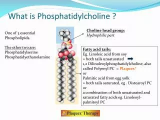
Abstract DNA demethylation by Ten-eleven translocation 1 (Tet1) has been shown to have an important role in differentiation and reprogramming of stem cells though the role of Tet1 in adult stem cells and cellular metabolism is still less understood. In our present study we used C2C12 mouse skeletal muscle precursor cells to test the role of Tet1 in regulation of metabolism. First we optimized the experimental conditions for siRNA knockdown of Tet1 expression in C2C12 cells using different lipid based transfection reagents. We observed a successful knockdown of Tet1 using the Lipofectamine (2000 and RNAiMAX) transfection reagents for siRNA treatment. By using a metabolic approach we assessed the effect of Tet1 knockdown on major cellular metabolic pathways. Our metabolomics and qRT-PCR results indicate that Tet1 significantly modulates glycerphosphocholine levels and alters expression of genes (Pcyt1a) involved in phosphatidylcholine biosynthesis. Similarly, silencing of Tet1 expression also significantly altered glycerphosphocholine levels in R1 mouse Embryonic Stem cells. We also observed that cells treated with a 60μM dose of phosphatidylcholine showed significant morphological changes and loss of alkaline phosphatase staining as compared to the untreated controls. All together our results indicate that Tet1 might have an important role in regulating phosphatidylcholine biosynthesis in C2C12 and R1 ES cells, implicating that epigenetic regulators have a key role in regulating cellular metabolism. Evaluating the Role of the DNA Demethylase Tet1 in Regulating Metabolism of Mouse Myoblast C2C12 Cells and R1 Embryonic Stem Cells Choline Choline Kinase-a, b siRNA transfection Zoe Respondek, Samiha Mateen, Sonnet Davis, Kylie Mitchell, Arvind Ramanathan The Buck Institute for Age Research, Novato, CA Research Experience for Undergraduates Fellowship Cell Culture Phosphocholine Pcyt 1a, b Aqueous CDP- choline Lipid CPT Phosphatidylcholine Intracellular Extraction of Metabolites LC- Mass Spectrometer Introduction Results • Ten-eleven translocation 1 (Tet1) is a member of the Tet protein family of DNA demethylases. DNA demethylation is a type of modification, which occurs at the 5-methylcytosine (5mC) position where the Tet enzymes helps in the oxidation of the methyl group to form 5-hydroxymethylcytosine (5hmC)¹ (Fig1). Tet1 is known to play an important role in reprogramming and differentiation of stem cells². • The objective of our present study is to investigate the role of Tet1 in regulating cellular metabolism utilizing C2C12 mouse myoblast cells. C2C12 cells are maintained as myoblasts through subculture and can be differentiated upon fusion of myoblasts into myotubes (Fig 2). • We also tested the role of Tet1 in R1 mouse embryonic stem cells which are maintained in their pluripotent state and can be differentiated into one of the three germ layers: endoderm, mesoderm, or ectoderm. The primary goal of this study was to explore the role of Tet1 in stem cell metabolism which is less understood. Regulation of Phosphatidylcholine Pathway by Tet1 in R1 ES Cells A. B. Glycerphosphocholines * p=0.00237 C. D. Figure 5: Results for R1 ES cells A. Knockdown of Tet1 gene expression by siRNA transfection using DharmaFECT1 B. LC-MS results for glycerphosphocholine metabolites of Tet1 knockdown compared to the control treatment group. C. The genes of phosphatidylcholine biosynthesis pathway4. D. RT-qPCR results for Pcytla expression in Tet1 knockdown.*Statistically significant metabolites p ≤ 0.05 p=0.087 A. Unmethylated CpG Methylated CpG Myoblast Methods A. C. B. Tet Histone A similar experiment was performed in R1 ES cells under identical treatment conditions. Protein estimations for the different samples was performed for the protein normalization of samples using a Bradford Coomassie Protein assay. Phosphatidylcholine Treatment and Alkaline Phosphatase Staining R1 ES cells were plated on a 24-well plate and treated the next day with control (0.1% chloroform), 30 μM or 60 μM 1-palmitoyl-2-arachidonyl-sn-glycero-3-phosphocholine in 0.1% chloroform for six days. The cells were then stained for the detection of alkaline phosphatase. The loss of alkaline phosphatase is a marker for differentiation of ES cells. Myotube DNA B. Alkaline Phosphatase Staining of Phosphatidylcholine Treated R1 ES Cells 5mC 5hmC Control (.1% CHCl3) C. Myofiber 30 μM PC 60 μM PC Figure 3: Knockdown of Tet1 expression in C2C12 cells with different transfection reagents A. 48 hour siRNA with DharmaFECT1 transfection reagent. B. 48 hour siRNA with Lipofectamine 2000 C. 72 hour siRNA with RNAiMAX. NS Not statistically significant p>0.05 10x Williams, et al. 2012; EMBO Reports. Siminiak, et al. 2005; European Heart Journal. Results Figure 1: Tet1 mediated DNA Demethylation A. CpG islands of the DNA can exist in either methylated or demethylated states. B. Structure of Tet enzymes: Tet1, Tet2 and Tet3. C. Mechanism of Tet mediated demethylation2. Figure 2: Myogenesis of Skeletal Muscle Cells Myoblasts align and differentiate into long myotubes (C2C12 cells pictured 10x), which then mature into full myofiber. Optimization of Tet1 Knockdown in C2C12 Cells NS Magnified Aims p=0.0024 Aim 1: Optimize experimental conditions for knocking down Tet1 expression in C2C12 cells using siRNA p=0.0193 Figure 6: Alkaline Phosphatase Staining of Phosphatidylcholine (PC) treated cells Red color staining in the cells indicates presence of alkaline phosphatase. The loss of alkaline phosphatase is a marker for differentiation and loss of pluripotency in ES cells. Aim 2: Define the metabolic pathways modulated by Tet1 in C2C12 cells Aim 3: Investigate the role of Tet1 in regulating metabolism of mouse C2C12 and R1 embryonic stem (ES) cells Conclusions Successfully optimized knockdown of Tet1 expression with siRNA in C2C12 cells using Lipofectamine (2000 and RNAiMAX) transfection reagents LC-Mass Spectrometry Based Metabolic Profiling in C2C12 Cells Methods Cell Culture A. C2C12 Cell Culture The C2C12 cells (early passages 7-18) were maintained in an incubator at 5% CO2;37ºC in DMEM media (supplemented with 10% FBS and 1% Penicillin Streptomycin). The media was changed every 2 days and the cells were split every 3 days at 50% confluence. B. R1 mouse ES cell Culture The R1 ES cells were cultured on 6-well coated plates with Matrigel using R1 ES cell media and maintained in an incubator at 5% CO2; 37ºC culture conditions. siRNA for Tet1 Knockdown Cells were plated at 40-45% confluency and treated the next day with 100nM control siRNA or Tet1 siRNA using different transfection reagents (DharmaFECT1, Lipofectamine 2000, and RNAiMAX). After 48-72 hours, the cell pellets were collected then processed either for RNA or protein analysis. A. RNA Extraction The RNA was extracted using the Qiagen RNeasy Mini-kit and then the concentrations of RNA were determined using Nanodrop. B. cDNA Synthesis Reverse transcription using iScript Reverse Transcription Supermix for RT-qPCR was done in a thermal cycler to obtain the cDNA. C. Real Time Quantitative PCR RT-qPCR with SYBR green was done with specific gene primers in the CFX to detect changes in gene expression between treatment groups. Metabolomics After 48 hours of siRNA treatment, an intracellular lipid extraction was performed on the C2C12 cells using methanol: chloroform extraction procedure. The lipid and aqueous samples were run using a Liquid Chromatography Mass Spectrometry based system with an Agilent 1200 LC and Agilent6520 QTOF-MS to target and find metabolites. The metabolomics of C2C12 and R1 ES cells indicate that phosphatidylcholine metabolism is regulated by Tet1 A. Tet1 might regulate transcription of enzymes in the phosphatidylcholine biosynthesis pathway Future Directions We would like to further optimize the Tet1 knockdown and repeat metabolomics with LC-MS in C2C12 and R1 ES cells Glycolysis B. Glycerphosphocholines C. Investigate whether Tet1 directly regulates phosphatidylcholine metabolism and its effect on differentiation in the two cell lines * * * * References Tan, et al. 2012; Tet family proteins and 5-hydroxymethylcytosine in development and disease. Development Williams, et al. 2012; Role of TET proteins in DNA methylation. EMBO Reports Siminiak, et al. 2005; Percutaneous trans-coronary-venous transplantation of autologous skeletal myoblasts in the treatment of post-infarction myocardial contractility impairment: the POZNAN trial. European Heart Journal Vance, et al. 2009; Physiological consequences of disruption of mammalian phospholipid biosynthetic genes. Journal of Lipid Research TCA D. Figure 4: LC-MS Metabolomics for C2C12 cells A. Procedure for obtaining intracellular metabolite data through LC Mass Spectrometry. B. LC-MS results show glycerphosphocholine related metabolites found after Tet1 knockdown. C/D. Glycolysis and Tricarboxylic Acid (TCA) metabolites were not altered by decreased Tet1 expression in C2C12 cells. *Statistically significant metabolites p ≤ 0.05 Acknowledgments We would like to acknowledge the Buck Institute for Research on Aging and the Lamba Lab. I would also like to recognize the REU Fellowship Program and Dr. Kristen Gates.