Download
1 / 94
940 likes | 1.07k Views
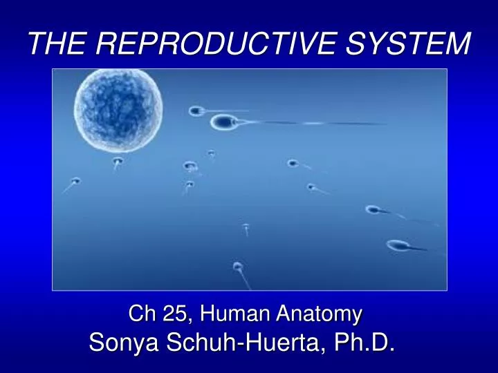
THE REPRODUCTIVE SYSTEM Ch 25, Human Anatomy Sonya Schuh-Huerta, Ph.D. The Reproductive System. Involved in the production of gametes, fertilization & procreation of the species Primary sex organs: Testes Ovaries Accessory sex organs: Glands External genitalia.

E N D
THE REPRODUCTIVE SYSTEM Ch 25, Human Anatomy Sonya Schuh-Huerta, Ph.D.
The Reproductive System • Involved in the production of gametes, fertilization & procreation of the species • Primary sex organs: • Testes • Ovaries • Accessory sex organs: • Glands • External genitalia
The Male Reproductive System • Produce male gamete (= sperm) • Testes are located within the scrotum • The scrotum Skin & superficial fascia surrounding the testes • Positioning provides an environment 3˚ cooler than body temperature! • Dartos muscle = layer of smooth muscle • Cremaster muscle = bands of skeletal muscle surrounding the testes • Elevates the testes
The Testes • Contain long coiled seminiferous tubules where • sperm are made • Enclosed in a serous sac the tunica vaginalis • Tunica albuginea fibrous capsule of the testes • Divides each testis into 250–300 lobules • Lobules contain 1–4 coiled seminiferous tubules • Epididymis • Comma-shaped structure on posterior testis; where the sperm go after they have developed in the testis
The Male Reproductive System Ureter Peritoneum Urinary bladder Seminal vesicle Prostatic urethra Ampulla of ductus deferens Pubis Ejaculatory duct Membranous urethra Rectum Urogenital diaphragm Prostate Corpus cavernosum Bulbourethral gland Corpus spongiosum Anus Spongy urethra Bulb of penis Ductus (vas) deferens Epididymis Glans penis Testis Prepuce (foreskin) Scrotum External urethral orifice
Relationship of the Testes to the Scrotum Urinary bladder Superficial inguinal ring (end of inguinal canal) Testicular artery Spermatic cord Ductus (vas) deferens Autonomic nerve fibers Penis Middle septum of scrotum Pampiniform venous plexus Cremaster muscle Epididymis External spermatic fascia Tunica vaginalis (from peritoneum) Superficial fascia containing dartos muscle Tunica albuginea of testis Scrotum Internal spermatic fascia Skin
Nerves & Vessels • Arterial supply of the testes • Testicular arteries • Testicular veins arise from the pampiniform plexus • Pampiniform plexus helps keep testes cool! • Testes innervated by parasympathetic & sympathetic divisions of ANS • The spermatic cord contains: • Vas deferens • Testicular blood vessels & nerves • Runs through inguinal canal
The Testis & Spermatic Cord Spermatic cord Spermatic cord Blood vessels and nerves Ductus (Vas) deferens Epididymis Testis Testis Head of epididymis Seminiferous tubule Efferent ductule Rete testis Lobule Straight tubule Septum Tunica albuginea Body of epididymis Tunica vaginalis Duct of epididymis Cavity of tunica vaginalis Tail of epididymis
Microscopic Anatomy • Scientists once believed that the sperm contained a tiny human or all the material that would give rise to the human (“spermists”)
A Seminiferous Tubule of the Testis Seminiferous tubule Spermatogenic cells Areolar connective tissue Sertoli Cell & nucleus Interstitial Cells (Leydig cells) Sperm Myoid cells
Microscopic Anatomy • Sertoli Cells • Surround developing sperm • Extend from basal lamina to the lumen • Tight junctions between cells • Blood-testis barrier (protects sperm from immune system!) • Assist sperm production • Secrete testicular fluid&androgen-binding protein (ABP)
Microscopic Anatomy • Interstitial cells: • Myoid cells surround seminiferous tubules • Contract rhythmically • Leydig cells • Secrete testosterone (T)! What does T do? • T secretion regulated by LH (Luteinizing Hormone)
The Epididymis • Duct of the epididymis is 6 m long (when uncoiled) • Dominated by pseudostratified columnar epithelium • Bears tufts of stereocilia immotile, long microvilli • 20-day journey for sperm to move through epidid. • Chemical changes that give them the ability to swim, undergo the acrosome reaction & fertilize an egg
The Epididymis Spermatic cord Blood vessels and nerves Ductus (Vas) deferens Testis Head of epididymis Seminiferous tubule Efferent ductule Rete testis Lobule Straight tubule Septum Tunica albuginea Body of epididymis Tunica vaginalis Duct of epididymis Cavity of tunica vaginalis Tail of epididymis
The Epididymis & Vas Deferens Smooth muscle around the duct of the epididymis Loose connective tissue outside the duct Stereocilia Duct of the epididymis Sperm in the lumen Pseudostratified columnar epithelium (a) Duct of the epididymis (110) Lumen Smooth muscle Mucosa Internal longitudinal layer Pseudostratified columnar epithelium Middle circular layer Lamina propria External longitudinal layer Adventitia (connective tissue) (b) Vas deferens (7)
Vas Deferens (Ductus Deferens) • Stores & transports sperm • Joins up with ejaculatory duct
The Vas Deferens • Trace the course of the Vas deferens Seminal vesicle Ampulla of ductus deferens Ejaculatory duct Bulbourethral gland Vas deferens
The Vas Deferens & Urethra Ureter Ampulla of ductus deferens Seminal vesicle Urinary bladder Ejaculatory duct Prostate Prostatic urethra Bulbourethral gland & duct Orifices of prostatic ducts Urogenital diaphragm Membranous urethra Bulb of penis Root of penis Crus of penis Bulbourethral duct opening Ductus deferens Corpora cavernosa Epididymis Corpus spongiosum Shaft (body) of penis Testis Spongy urethra Prepuce (foreskin) Glans penis External urethral orifice
Accessory Glands • The Seminal vesicles • Lie on the posterior surface of the urinary bladder • Secretes about 60% of the volume of semen • Fluid contains • Fructose to nourish sperm • Substances that enhance sperm motility • Prostaglandins • Substances that suppress immune responses against semen • Enzymes that clot & then liquefy semen
Accessory Glands • The Prostate gland • Encircles the prostatic urethra • Secretes about 25–30% of the volume of seminal fluid • Contains substances that • Enhance sperm motility • Enzymes that clot & then liquefy semen
Accessory Glands • The Bulbourethral glands • Pea-sized glands inferior to the prostate gland • Produce a mucus • Mucus enters spongy urethra prior to ejaculation • Neutralizes traces of acidic urine, otherwise this would kill sperm! • Also lubricates urethra
Male External Genitalia • Penis internal anatomy • 3 erectile bodies • 1 corpus spongiosum • Surrounds spongy urethra • 2 coropora cavernosa • Contain sinuses, make up most of the penis • Male sexual response • Erection parasympathetic control, allows blood to enter corpora cavernosa • Ejaculation sympathetic control
Spermatogenesis • Sperm formation • 400 million sperm formed per day (that’s ~4,000 sperm per heartbeat!) • Begins at puberty, continues throughout man’s entire life • Process takes ~75 days • Cells undergo meiosis, differentiate, & move toward the lumen • Spermatogonia • Primary spermatocytes • Secondary spermatocytes • Spermatids • Sperm 1° spermatocyte 2° spermatocyte TESTIS mature sperm spermatogonium spermatid gonocyte
Spermatogenesis • Divided into 3 stages • Stage 1 spermatogonia divide by mitosis • Type A maintain the germ cells (= stem cells) • Type B differentiate into primary spermatocytes • Stage 2 meiosis I & meiosis II • Meiosis I forms 2 secondary spermatocytes • Meiosis II each secondary spermatocyte forms 2 spermatids • Stage 3 Spermiogenesis • The 4 spermatids differentiate into mature sperm • Flagellum (tail) formed ( head, midpiece, tail) • Form acrosome • Shed cytoplasm (compact head, with very little cytosol)
Spermatogenesis Basal lamina Spermatogonium (stem cell) Type A daughter cell remains at basal lamina as a stem cell Mitosis Growth Type B daughter cell Enters meiosis I and moves to adluminal compartment Primary spermatocyte Meiosis (early spermatogenesis) Meiosis I completed Secondary spermatocytes Meiosis II Spermatogenesis Early spermatids Late spermatids Spermiogenesis (late spermatogenesis) Sperm (b) Events of spermatogenesis, showing the relative position of various spermatogenic cells
Spermiogenesis Approximately 24 days Golgi apparatus Acrosomal vesicle Mitochondria Acrosome Nucleus 1 2 Spermatid nucleus Centrioles Midpiece Head Microtubules 3 (a) Flagellum Excess Cytoplasm (cytoplasmic droplet) Acrosome 4 Tail 5 7 6 (b)
Control of Spermatogenesis • Spermatogenesis controlled by • Follicle stimulating hormone = FSH (from ant. pituitary) • Testosterone (from testis) • Secretions from Sertoli cells also influence spermatogenesis • Androgen-bindingprotein concentrates T near developing sperm • Inhibin inhibits FSH (feedback loop)
Normal Sperm Parameters & Male Infertility • > 20 million (20–150 million) sperm per ml (2–3 ml/ejaculate) • > 50% forward motility • > 40% normal morphology Male Infertility Disorders • Endocrine – Testicular Feminization Syndrome (androgen receptor does not respond to T) • Hypothalamic – Kallman’s Syn (no GnRH or olf. neurons) • Anatomic – Varicocele • Sertoli Cell Only Syndrome • Oligospermia = reduced sperm # • Azoospermia = no sperm • Asthenozoospermia = reduced motility • Chromosomal Defects = Klinefelter’s Syndrome (XXY) human sperm
Causes of Human Infertility • ~15% of couples are infertile • Male infertility contributes to ~½ of cases low sperm quantity poor morphology poor motility Causes of Male Infertility • Endocrine • Hypothalamic/Pituitary • Anatomic • Coital Disorders • Environmental Factors • Infections/toxins/drugs • Sperm abnormalities • Genetic/Chromosomal Defects • Idiopathic Ovulatory Dysfunction 15% Unusual Problems 5% Female tubal & pelvic pathology 35% Unknown 10% Male Factor 35% (Speroff, L., Clin Gynecol Endocrin & Infertility, 1999)
Cancers of the Male Reproductive System • Testicular cancer • Affects 1 in 50,000 males • Commonly from early-stage spermatogenic cells • Increase of 50% from 1974 to 1990 • Cured in 95% of cases • Prostate cancer • Slow-growing arises from peripheral glands • Increasingly common • Risk factors • Fatty diet • Genetic predisposition
The Female Reproductive System Leonardo Da Vinci
Female Reproductive System • Produces female gamete (= oocytes/eggs) • Functions to support a developing embryo • Undergoes changes according to the menstrual cycle • Menstrual cycle = the monthly cycle as it affects all reproductive organs in preparation for pregnancy • Includes: • Ovaries, uterine tubes (fallopian tubes), uterus, & vagina
The Vagina • Consists of 3 layers • Adventitia fibrous connective tissue • Muscularis smooth muscle • Mucosa marked by transverse folds • Consists of lamina propria & stratified squamous epithelium • Hymen an incomplete diaphragm • Fornix recess formed at the superior part of the vagina
The Ovaries • Small, almond-shaped organs • Produce eggs/oocytes!!! • Held in place by ligaments & mesenteries • Broad, suspensory, & ovarian ligaments • Ovarian arteries arterial supply • Innervated by both divisions of ANS
Female Internal Reproductive Organs Suspensory ligament of ovary Infundibulum Uterine tube Ovary Peritoneum Fimbriae Uterus Uterosacral ligament Round ligament Perimetrium Vesicouterine pouch Rectouterine pouch Urinary bladder Pubic symphysis Rectum Mons pubis Posterior fornix Urethra Cervix Clitoris Anterior fornix External urethral orifice Vagina Anus Hymen Urogenital diaphragm Labium minus Greater vestibular (Bartholin’s) gland Labium majus
The Female Reproductive System Suspensory ligament of ovary Uterine (fallopian) tube Uterine tube Ovarian blood vessels Fundus of uterus Lumen (cavity) of uterus Ampulla Ovary Mesosalpinx Isthmus Infundibulum Mesovarium Broad ligament Fimbriae Mesometrium Round ligament of uterus Ovarian ligament Endometrium Body of uterus Myometrium Wall of uterus Perimetrium Ureter Internal os Uterine blood vessels Cervical canal Isthmus External os Uterosacral ligament Lateral cervical (cardinal) ligament Lateral fornix Vagina Cervix
Deep Structures of the Female External Genitalia & Perineum Clitoris Labia minora Labia majora Anus Pubic symphysis Body of clitoris, containing corpora cavernosa Inferior ramus of pubis Clitoris (glans) Crus of clitoris Urethral orifice Vaginal orifice Bulb of vestibule Greater vestibular gland Fourchette
Internal Structure of the Ovaries follicle B oocyte • Tunica albuginea • Fibrous capsule of the ovary • Ovarian cortex periphery, where developing oocytes are found • Follicles multi-cellular sacs housing oocytes • Ovarian medulla loose connective tissue • Contains blood vessels, lymph vessels, & nerves
Structure of the Ovary Tunica albuginea Granulosa cells Cortex Antral follicle Degenerating corpus luteum (corpus albicans) Oocyte Mesovarium & blood vessels Germinal epithelium Mature ovarian (Graafian) follicle Primary follicles Antrum Oocyte Ovarian ligament Zona pellucida Theca folliculi Medulla Ovulated oocyte Corpus luteum Corona radiata Developing corpus luteum
The Fallopian Tubes (Oviducts) • Receive ovulated oocyte • Parts of the fallopian tube • Infundibulum = distal end of uterine tube • Surrounded by fimbriae (finger-like projections) • Ampulla = middle 1/3 of uterine tube • Usual site of fertilization!!! • Isthmus = medial 1/3 of uterine tube; narrows
Microscopic Anatomy of Uterine Tubes • Muscular & ciliated aids in transport of oocyte & embryo! Muscularis Ciliated columnar epithelium Mucosa Nonciliated epithelium Lamina propria (a) Cross section through the ampulla (10) (b) Enlargement of the mucosa (180)
The Uterus • Lies anterior to rectum, posterior to bladder • Anteverted (anteflexed) usual position of uterus • Parts of the uterus • Fundus = rounded superior portion • Cervix = “neck” of uterus • Cervical canal = connects with vagina inferiorly • Internal os = opening connecting with uterine cavity • External os = inferior opening of cervix (at top of vagina)
Supports of the Uterus • Uterus is supported by • Mesometrium anchors uterus to lateral pelvic walls • Lateral ligaments horizontal from cervix & vagina • Round ligaments bind uterus to the anterior pelvic wall
Uterine Wall • Wall of the uterus composed of: • Perimetrium, myometrium, & endometrium • Embryo burrows into endometrium • Endometrium has 2 layers • Functional layer • Basal layer
Uterine Wall • Uterine arteries send branches to uterine wall & divide into arcuate arteries • Radial arteries reach the endometrium & branch into • Straight arteries to basal layer • Spiral arteries to functional layer • Undergo degeneration (& are shed) and then regeneration with the menstrual cycle
The Uterine Wall Lumen of uterus Lumen of uterus Epithelium Epithelium Capillaries Uterine glands Uterine glands Venous sinusoids Lamina propria of connective tissue Functional layer of the endometrium Lamina propria of connective tissue Spiral (coiled) artery Straight artery Endometrial vein Smooth muscle fibers Basal layer of the endometrium Radial artery Smooth muscle fibers Arcuate artery Portion of the myometrium Radial artery Uterine artery
The Female Menstrual Cycle & Oogenesis • Monthly menstrual cycle • The recurring cycle of physiological changes to uterus, ovaries, & other reproductive organs due to hormonal fluctuations in ovaries, that occur from the beginning of one menstrual period to the next • Ovarian cycle stimulates maturation of ovarian follicles & oocytes • Uterine cycle prepares uterine wall for implantation
The Ovarian Cycle • Follicular phase • 1st half of the ovarian cycle • 6–12 primordial follicles stimulated to develop • Growth stimulated by FSH from ant. pituitary • Primordial follicle primary follicle • As follicles grow larger they gain granulosa & thecal cells layers of follicle that secrete hormones (estrogens, etc.)
The Follicular Phase (cont…) • Primary follicles secondary follicles • Corona radiata coat of granulosa cells surrounding oocyte • Secondary follicles enlarge & become • Antral follicles (Antrum = fluid-filled cavity) • Then,become mature Graafianfollicle ready to be ovulated • Only 1 dominant Graafian follicle ovulated (usually!) • Rest undergo atresia cell death Graafian follicle Antral follicle OVARY 1° follicle 2° follicle primordial follicle mature ovum gonocyte ovulation