Download
1 / 54
540 likes | 544 Views
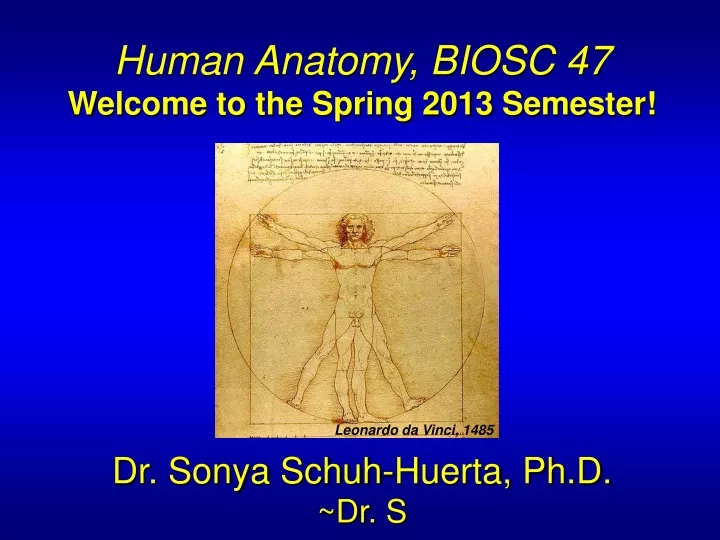
Human Anatomy, BIOSC 47 Welcome to the Spring 2013 Semester! Dr. Sonya Schuh-Huerta, Ph.D. ~Dr. S. Leonardo da Vinci, 1485. Who am I?…. Research Scientist at Stanford & Part-time Assoc. Prof. at Mission College

E N D
Human Anatomy, BIOSC 47 Welcome to the Spring 2013 Semester! Dr. Sonya Schuh-Huerta, Ph.D. ~Dr. S Leonardo da Vinci, 1485
Who am I?… • Research Scientist at Stanford & Part-time Assoc. Prof. at Mission College • Earned my Ph.D. in Physiology & Biophysics from the Univ. of Washington (Seattle); before that HSU; before that UCR; before that – a small community college in Southern California… • My research is on human development, reproductive biology and genetics, fertility, & stem cell biology • I love teaching! • I expect a lot from myself & from my students – this class will be challenging, but we’ll also have a lot of fun! • One word of advise – hard work is more important than intelligence, innate ability, or anything else. With hard-work & perseverance you can truly conquer any goal. • On a personal note – I have 3 kids, 1 husband, 2 dogs, & 2 cats..
What is Anatomy? • Anatomy • The study of the structure of the body • Physiology • The study of body function • Anatomy & Physiology are closely related & you need to understand a bit about both as you are learning them
Overview of Anatomy • Branches of anatomy • Gross Anatomy • Microscopic Anatomy (Histology) • Surface Anatomy • Developmental Anatomy • Embryology • Pathological Anatomy (Pathology) • Radiographic Anatomy • Functional Morphology • Anatomical terminology • Based on ancient Greek or Latin • Provides standard nomenclature worldwide
The Hierarchy of Structural Organization • Chemical level – atoms form molecules • Cellular level – cells and their functional subunits • Tissue level – a group of cells performing a common function • Organ level – a group of different types of tissues working together
The Hierarchy of Structural Organization Organelle Molecule Atoms Smooth muscle cell 2 Cellular level Cells are made up of molecules. 1 Chemical level Atoms combine to form molecules. Smooth muscle tissue Cardiovascular system 3 Tissue level Tissues consist of similar types of cells Heart Blood vessel (organ) Blood vessels Smooth muscle tissue Connective tissue Epithelial tissue 4 Organ level Organs are made up of different types of tissues. 5 Organ system level Organ systems consist of different organs that work together closely. 6 Organismal level The human organism is made up of many organ systems.
Integumentary System • Forms external body • covering • Protects deeper tissues • from injury • Synthesizes vitamin D • Site of cutaneous receptors • -pain, pressure, etc. & • sweat & oil glands Hair Nails Skin
Skeletal System • Protects & supports body • organs • Provides a framework for • muscles • Blood cells formed within • bones • Stores minerals (calcium) Bones Joint
Muscular System • Allows manipulation of • environment • Locomotion • Facial expression • Maintains posture • Produces heat Skeletal muscles
Nervous System • Fast-acting control system • Controls many body • functions • Responds to internal & • external changes Brain Nerves Spinal cord
Endocrine System Pineal gland • Involved in many processes; • glands secrete hormones • that regulate: • Growth • Reproduction • Metabolism • Circadian rhythms Pituitary gland Thyroid gland Thymus Adrenal gland Pancreas Testis Ovary
Cardiovascular System • Blood vessels transport blood • Carries O2 & CO2 • Carries nutrients & wastes • Heart pumps blood through • blood vessels Heart Blood vessels
Lymphatic / Immune System • Picks up fluid leaked from blood • vessels • Disposes of debris • Houses white blood cells • Mounts attack against foreign • substances in the body Red bone marrow Thymus Lymphatic vessels Thoracic duct Spleen Lymph nodes
Respiratory System • Keeps blood supplied w/ O2 • Removes CO2 • Gas exchange occurs through • walls of air sacs • (alveoli in lungs) Nasal cavity Pharynx Bronchus Larynx Trachea Lung
Digestive System • Breaks down food into • absorbable units • Indigestible foodstuffs • eliminated as feces • Also secretes hormones • involved in appetite & • metabolism Oral cavity Esophagus Liver Stomach Small intestine Large intestine Rectum Anus
Urinary System • Eliminates nitrogenous • wastes as urine • Regulates water, electrolyte, • & acid-base balance Kidney Ureter Urinary bladder Urethra
Reproductive System • Overall function = produce offspring • Testes produce sperm & male sex hormones • Ovaries produce eggs & female sex hormones • Mammary glands produce milk Mammary glands (in breasts) Prostate gland Ovary Penis Ductus deferens Testis Uterine tube Scrotum Uterus Vagina
Scale: Length, Volume, & Weight • Metric system = provides a precise system of measurement • Weight (mass) grams (g), kilograms (kg) • Volume liters (l), milliliters (ml) • Length meters (m), centimeters (cm), micrometers (mm)… • -Average adult = 1.5 – 2.0 meters long • -Cells & tissues are measured in mm • -Avg cell diameter = 10 mm • -Largest cell oocyte! (~100+ mm) oocyte within follicle
Gross Anatomy – An Introduction • Anatomical position – a common visual reference point • Person stands erect with feet together & eyes forward • Palms face anteriorly with thumbs pointed away from body • Directional terminology – refers to the body in anatomical position • Standardized terms of directions are paired terms
Gross Anatomy – An Introduction • Directional terms • Regional terms = names of specific body areas • Axial region = the main axis of the body • Appendicular region = the limbs
Regional Terms of Gross Anatomy Axial region Appendicular region Cephalic (head) Frontal Upper limb Orbital Acromial Nasal Brachial (arm) Oral Antecubital Mental Cervical (neck) Antebrachial (forearm) Thoracic Axillary Carpal (wrist) Sternal Mammary Manus (hand) Abdominal Pollex Umbilical Palmar Pelvic Digital Inguinal (groin) Lower limb Coxal (hip) Femoral (thigh) Patellar Pubic (genital) Crural (leg) Fibular or peroneal Pedal (foot) Thorax Tarsal (ankle) Abdomen Metatarsal Back (Dorsum) Digital Hallux (a) Anterior/Ventral
Regional Terms of Gross Anatomy Cephalic Appendicular region Otic Occipital (back of head) Upper limb Acromial Cervical Brachial (arm) Back (dorsal) Olecranal Scapular Antebrachial (forearm) Vertebral Lumbar Manus (hand) Sacral Metacarpal Gluteal Digital Perineal (between anus and external genitalia Lower limb Femoral (thigh) Popliteal Sural (calf) Fibular or peroneal Thorax Pedal (foot) Abdomen Calcaneal Back (Dorsum) Plantar (b) Posterior/Dorsal
Body Planes and Sections • Coronal (frontal) plane = Lies vertically & divides body into anterior & posterior parts • Median (midsagittal) plane = Specific sagittal plane that lies vertically in the midline • Transverse plane = Runs horizontally & divides body into superior & • inferior parts Frontal plane Median plane (midsagittal) Transverse plane
Characteristics Common to All Vertebrates • Tube-within-a-tube • Bilateral symmetry • Dorsal hollow nerve cord • Notochord & vertebrae • Segmentation • Pharyngeal pouches
Basic Human Body Plan & Vertebrate Structures Notochord Muscle segments (myotomes) Brain Spinal cord Brain Pharynx Muscle segments (muscles between ribs) Pharyngeal pouches Digestive tube Heart (a) Generalized vertebrate Spinal cord Lung bud Pharyngeal pouches Heart Vertebrae Spinal cord Disc between vertebrae Notochord Digestive tube Muscle segments (myotomes) (c) Adult human Digestive tube Heart Brain Inner tube Segmented outer tube (b) Human embryo; 5 weeks postconception Dorsal hollow nerve tube Notochord
Body Cavities & Membranes Cranial cavity (contains brain • Dorsal body cavity • Cranial cavity • Vertebral cavity Dorsal body cavity Thoracic cavity (contains heart and lungs) Vertebral cavity (contains spinal cord) Diaphragm Abdominal cavity (contains digestive viscera) Pelvic cavity (contains urinary bladder, reproductive organs, and rectum) Dorsal body cavity Ventral body cavity (a) Lateral view
Body Cavities & Membranes • Ventral body cavity • Thoracic cavity – divided into 3 parts • Two lateral parts each containing a lung surrounded by a pleural cavity • Mediastinum – contains the heart surrounded by the pericardial sac • Abdominopelvic cavity – divided into 2 parts • Abdominal cavity – contains the liver, stomach, kidneys, and other organs • Pelvic cavity – contains the bladder, some reproductive organs, & rectum
Ventral Cavities Cranial cavity Dorsal body cavity Ventral body cavity Vertebral cavity Superior mediastinum Thoracic cavity (contains heart and lungs) Pleural cavity Pericardial cavity within the mediastinum Ventral body cavity (thoracic and abdominopelvic cavities) Diaphragm Abdominal cavity (contains digestive viscera) Abdomino- pelvic cavity Pelvic cavity (contains urinary bladder, reproductive organs, and rectum) (b) Anterior view
Body Cavities & Membranes • Serous cavities – a slit-like space lined by a serous membrane • Pleura, pericardium, & peritoneum • Parietal serosa – outer wall of the cavity • Visceral serosa – inner wall of the cavity; covers the visceral organs
Body Cavities & Membranes Outer balloon wall (comparable to parietal serosa) Air (comparable to serous cavity) But no air in serous cavities! Inner balloon wall (comparable to visceral serosa) Model of the serous membranes & serous cavities
Body Cavities & Membranes Lung Ribs Parietal pleura Pleural cavity with serous fluid Visceral pleura Diaphragm Serosae associated with the lungs: pleura
Body Cavities & Membranes Heart Parietal pericardium Pericardial cavity with serous fluid Visceral pericardium Serosae associated with the heart: pericardium
Body Cavities & Membranes Visceral peritoneum Anterior Peritoneal cavity (with serous fluid) Liver Stomach Parietal peritoneum Kidney (retroperitoneal) Wall of body trunk Posterior Serosae associated with the abdominal viscera: peritoneum Figure 1.7c
Abdominal Regions & Quadrants • Abdominal regions divide abdomen into 9 regions • Abdominal quadrants divide abdomen into 4 • quadrants • Right upper & left upper quadrants • Right lower & left lower quadrants
Right hypochondriac region Left hypochondriac region Epigastric region Right lumbar region Left lumbar region Umbilical region Right iliac (inguinal) region Hypogastric (pubic) region Left iliac (inguinal) region (a) 9 regions delineated by 4 planes Abdominal Regions Diaphragm Liver Spleen Gallbladder Stomach Transverse colon of large intestine Ascending colon of large intestine Descending colon of large intestine Small intestine Cecum Initial part of sigmoid colon Appendix Urinary bladder (b) Anterior view of the nine regions showing thesuperficial organs
Abdominal Quadrants Right upper quadrant (RUQ) Left upper quadrant (LUQ) Right lower quadrant (RLQ) Left lower quadrant (LLQ) (c) The 4 abdominopelvic quadrants
Microscopic Anatomy • Microscopy – examining small structures through a microscope • Antonie van Leeuwenhoek • 1st discovered & examined cells • (“animalcules”) with homemade • microscopes in mid-1600s • -Light microscopy = illuminates tissue • with a beam of light (lower magnification) • -Transmission electron microscopy = • uses beam of electrons (higher mag); • specimens coated w/ heavy-metal salts, • which deflect electrons to different extents
Microscopic Anatomy • Scanning electron microscopy • Coat specimen with carbon & gold – when electron beam scans specimen, secondary electrons are emitted & detected beautiful 3D images assembled! • These images give amazing surface detail of cells & small structures. • Artifacts • Minor distortions of preserved tissues • Not exactly like living tissues & organs
Microscopic Anatomy Cytoplasm Cell nuclei Extracellular material (a) Light micrograph (330) (c) Scanning electron micrograph, artificially colored (2900) (b) Transmission electron micrograph, artificially colored (870)
Microscopic Anatomy • Preparing human tissue for microscopy • Specimen is fixed (preserved) & sectioned • Specimen is stained to distinguish structures • Acidic stain – negatively charged dye molecules • Basic stain – positively charged dye molecules
Clinical Anatomy – An Introduction to Medical Imaging Techniques • X ray – electromagnetic waves of very short length • Best for visualizing bones and abnormal dense structures Clavicles (collarbones) Ribs Air in lungs (black) Heart Diaphragm (a) Radiograph of the chest (b) Mammogram (cancerous tumor at arrow)
Advanced Imaging Techniques • Computed (axial) tomography(CT or CAT) = takes successive X rays around a person's full circumference • Creates detailed picture of body sections (transverse) • Great for soft tissue & bone; fast & inexpensive! Right Left Liver Stomach Colon View Inferior vena cava Aorta Spleen Left kidney Thoracic vertebra
Advanced Imaging Techniques • Angiography or digital subtraction angiography (DSA)imaging = provides an unobstructed view of small arteries • Contrast medium is injected • Used to identify blockages of arteries that supply heart or brain Artery supplying heart Narrowing of the artery