Download
1 / 1
10 likes | 94 Views
Explore the genetic and developmental aspects of INCL through PPT1 gene analysis in zebrafish embryos and adults. Determine spatial and temporal gene expression. Future experiments include morpholino knockdown. Literature cited supports small vertebrate models.

E N D
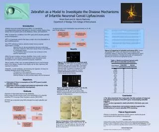
Zebrafish as a Model to Investigate the Disease Mechanisms of Infantile Neuronal Ceroid Lipfuscinosis Nicole Brant and Dr. Wendy Boehmler, Department of Biology, York College of Pennsylvania http://www.devbio.uga.edu/gallery/images/embryo4LRG.jpg http://www.bcm.edu/cain_foundation/noframes/html/pages/staff/neurons%20confocal%20mu1a%20dcx.jpg • Introduction • Infantile neuronal ceroid lipfuscinosis (INCL) is a devastating neurodegenerative disorder that destroys neurons in certain tissues of the central nervous system such as the thalamus, cortex, and cerebellum. • INCL is caused by a mutation in the PPT1 gene which encodes palmitoyl protein thioesterase 1. • PPT1 is a lysosomal enzyme that plays a major role in the degradation of lipid-modified proteins. • How PPT1 deficiency leads to selective central nervous system degeneration is unclear. • Neurons have an altered endoplasmic reticulum morphology. • Microarray analyses on INCL brain tissues shows an upregulation of inflammatory genes. • The post-mitotic nature of neurons may contribute to their selective degeneration. • While the gene mutation has been identified, there is still a need to investigate the neurobiology of the disease course throughout early development and to assess potential therapeutic treatments. • Mouse models of INCL have provided significant clues to the biological basis of the disease, however such studies can be slow, laborious, and expensive to perform. • Zebrafish, a small vertebrate, are becoming the organism of choice for modeling diseases and for drug discovery. • The genome is fully sequenced. • Breeding pairs can produce hundreds of embryos. • Embryos develop ex utero, develop rapidly, and are transparent • A whole-mount in situ hybridization was performed on 24, 48, and 72 hpf embryos. 1033 bp Washes Results Figure 4: Comparison of zebrafish and human PPT1. Human and zebrafish PPT1 amino acid sequences were aligned using CLUSTALW. Dashes in sequences allow optimal alignment for amino acid insertions/deletions. Identical amino acids are highlighted by asterisks and conserved are highlighted by dots. Ladder Brain Eye Gut Muscle Heart Table 1: Markers and Genes Syntenic with PPT1 Genes in Zebrafish and Humans. Figure 1: RT-PCR analysis of PPT1 from adult zebrafish. The PPT1 gene was amplified from adult zebrafish brain, gut, and muscle RNA. The primers used encompassed the predicted ATG start codon and stop codon to produce an amplicon of 1033 base pairs (bp). Primer sequences: forward 5’AGATTGAATAATGGCTCCACC3’ and reverse 5’TATCTGAGACGGTAGTTACGA3’. Ladder 24 hour 48 hour 72 hour Ladder • Objectives • Determine if there is expression of the PPT1 gene in adult zebrafish tissues. • Determine if there is spatial and temporal expression of the PPT1 gene during zebrafish development. • Methods • Searches of expressed sequence tag databases (http://www.ncbi.nlm.nih.gov/) led to the identification of a potential zebrafish PPT1 homolog (Gene Accession number NM213339). • RT-PCR was conducted using RNA collected from adult zebrafish and embryos. 1033 bp Figure 2: RT-PCR analysis of PPT1 from 24, 48, and 72 hpf embryos. The PPT1 gene was amplified from 24, 48, and 72 hpf embryo RNA. Primers encompassed the entire gene to produce the predicted 1033bp amplicon. • Conclusion • The data presented here represents the first analysis of temporal and spatial expression of the PPT1 gene in developing zebrafish embryos. • PPT1 is also expressed in adult zebrafish in the brain, gut, and muscle. • Sequence comparisons and syntenic analysis supports the identification of a novel zebrafish PPT1 homolog. • Future Experiments • Perform a morpholino knockdown of PPT1 gene to assess any gross morphological abnormalities in development. • Literature Cited • Cooper, Jonathan D., Russel, Claire and Mitchison, Hannah, M. 2006. Progress towards understanding disease mechanisms in small vertebrate models of neuronal ceroid lipofuscinosis. Biochimica et Biophysica Acta 873-889. • Woods, I. G., Wilson, C., Friedlander, B., Chang, P., Reyes, D. K., Nix, R., Kelly, P. D., Chu, F., Postlethwait, J. H., and W. S. Talbot. 2005. The zebrafish gene map defines ancestral vertebrate chromosomes. Genome Research 15:1307-1314. SC B E A 24 hpf D 24 hpf B SC E Collected tissues from the brain, eye, gut, heart, and muscle and 24, 48, and 72 hours post fertilization (hpf) embryos Collected 24, 48, and 72 hpf embryos, stored them in MeOH, made the sense and antisense probes and reagents RNA isolation with phenol/chloroform extraction B 48 hpf E 48 hpf Permeabilization of the embryos and hybridization of the RNA probe B SC E Reverse Transcription Washes PCR Denature for 30 sec. at 94oC Annealing for 30 sec. at 48oC Elongation for 1 min. at 72oC 30 cycles C 72 hpf F 72 hpf Figure 3: Expression of the palmitoyl protein thioesterase 1 gene. Lateral views of 24 (A, D), 48 (B, E), and 72 (C, F) hpf embryos. Note the sense probe (A, B, C) and the antisense probe (D, E, F). B-brain, E-eye, SC-spinal cord Incubation with anti-DIG antibody alkaline phosphase Staining and washing, stored embryos and took pictures cDNA Zebrafish PPT1 gene was successfully cloned into a pDrive and sequenced Acknowledgements I would like to thank Dr. Boehmler for all of her time and guidance.