Download

1 / 47
470 likes | 486 Views
Conception and Biophysical changes. Oogenesis. Ovary gives rise to oogonial cells First meiotic division – two unequal cells Second meiotic division – begins at ovulation Three polar bodies One ovum.

E N D
Oogenesis • Ovary gives rise to oogonial cells • First meiotic division – two unequal cells • Second meiotic division – begins at ovulation • Three polar bodies • One ovum
Figure 11-3-A Gametogenesis involves meiosis within the ovary and testis. During meiosis, each oogonium produces a single haploid ovum once some cytoplasm moves into the polar bodies.
Spermatogenesis • Production of sperm begins during puberty • First meiotic division • Primary spermatocyte replicates and divides • Second meiotic division • Secondary spermatocytes replicate and divide • Produce four spermatids • Series of changes
Figure 11-3-B Gametogenesis involves meiosis within the ovary and testis. Each spermatogonium, in contrast, produces four haploid spermatozoa.
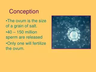
The Process of Fertilization • Sperm and ovum unite to form a zygote • Ova are fertile for 12 to 24 hours • Sperm are fertile for 72 hours • Takes place in the ampulla of fallopian tube
Conception • Necessary functional components • a. uterus • b. fallopian tubes • c. ovaries (eggs) • d. hormones • e. sperm
Preparation for Fertilization • Estrogen levels increase peristalsis in fallopian tubes • Fertilization usually takes place in ampulla • Single ejaculation – 200 to 500 million spermatozoa • Prostaglandins in semen help transport sperm
Changes in Sperm • Capacitation • Removal of plasma membrane and glycoprotein coat • Loss of seminal plasma proteins • Acrosomal reaction • Release of enzymes • Allows entry through corona radiata
After Sperm Entry • Additional sperm blocked by zone pellucida • Secondary oocyte completes second meiotic division, forms nucleus of ovum • Nuclei of ovum and sperm unite • Nuclear membranes disappear • Chromosomes pair up • Sex of zygote determined
Fertilization & implantation • Several Days Pass Until Bilaminar Blastocyst Implants (6 days) • b. double layer development • 1) blastocyst - inner layer - solid • 2) trophoblast - outer layer - foraging layer (feeding layer) • c. implantation • d. trophoblast to chorion
Figure 11-4 A. Sperm penetration of an ovum. The sequential steps of oocyte penetration by a sperm are depicted moving from top to bottom.
Figure 11-4 B. Sperm penetration of an ovum. Scanning electron micrograph of human sperm surrounding a human oocyte (750 ). The smaller spherical cells are granulosa cells of the corona radiata. Source: Used with permission from Nilsson, L. (1990). A child is born. New York, NY: Dell Publishing.
Preembryonic Stage • First 14 days of human development • Cleavage • Blastomeres form morula • Blastocyst • Develops into embryonic disc and amnion • Trophoblast • Develops into chorion
Implantation • Occurs 7 to 10 days after fertilization • Blastocyst burrows into endometrium • Endometrium is now called decidua
Cellular Differentiation • Primary germ layers • Ectoderm • Mesoderm • Endoderm • All tissues develop from primary germ cell layers
Table 11-2 Derivation of Body Structures from Primary Cell Layers
Cellular Differentiation (cont’d) • Embryonic membranes begin to form • Chorion(outerm membrane contribues to placental development) • Amnion (inner most membranous sac) • Amniotic fluid • Functions –protects against physical force, maintains temperature, allows fetal movement • Characteristics-clear, slightly yellow, 700-1000cc by 3rd trimester, • Abnormal variations- • Yolk sac-provides early nourshment
Cellular Differentiation (cont’d) • Umbilical cord • Body stalk fuses with embryonic portion of placenta • Provides circulatory pathway from chorionic villi to embryo • One vein, two arteries • Wharton’s jelly • Delivers oxygenated blood to fetus • Two arteries
Placenta • Metabolic and nutrient exchange • Maternal portion • Decidua basalis and circulation • Fetal portion • Covered by amnion (chorionic villi) • Fetal surface covered by amnion
Placental Development • Chorionic villi form spaces in decidua basalis • Spaces fill with maternal blood • Chorionic villi (provide blood supply to the embryo) • Syncytium: outer layer • Cytotrophoblast: inner layer • Anchoring villi form septa
Figure 11-13 Longitudinal section of placental villus. Spaces formed in the maternal decidua are filled with maternal blood; chorionic villi proliferate into these maternal blood-filled spaces and differentiate into a syncytium layer and a cytotrophoblast layer.
Placental Circulation • After implantation cells differentiate • Trophoblast invades endometrium • Opens uterine capillaries • Completion of maternal-placental-fetal circulation • About 17 days after conception
Figure 11-14 Vascular arrangement of the placenta. Arrows indicate the direction of blood flow. Maternal blood flows through the uterine arteries to the intervillous spaces of the placenta and returns through the uterine veins to maternal circulation. Fetal blood flows through the umbilical arteries into the villous capillaries of the placenta and returns through the umbilical vein to the fetal circulation.
Placental Facts • Size of 8-inch dinner plate • Consists of 15-20 cotyledons, chorionic villi branch out from it • Structure is complete at the end of 12th week gestation • Grows till 20th week gestation, covers ½ of uterine surface
Placental Functions • Transfers nutrients • Diffuses oxygen and carbon dioxide functioning as fetal lungs • Production of 4hormones • hCG • Progesterone • Estrogens • hCS/hPL • Facilitates transfer of metabolic wastes from the fetus to the maternal ciruclation • Transfers heat from mother to fetus.
Development of the Fetal Circulatory System • Maintains blood flow to placenta • Provides fetus with oxygen and nutrients • Removes carbon dioxide and waste products • Blood flows through umbilical vein into abdominal wall of fetus
Figure 11-15 Fetal circulation. Blood leaves the placenta and enters the fetus through the umbilical vein. After circulating through the fetus, the blood returns to the placenta through the umbilical arteries. The ductus venosus, the foramen ovale, and the ductus arteriosus allow the blood to bypass the fetal liver and lungs.
Embryonic and Fetal Development • Pregnancy averages 10 lunar months • Beginning of last normal menstrual period to birth • Most born within 10 to 14 days of calculated date of birth
Embryonic Stage • Day 15 to Week 8 • Tissues differentiate into essential organs • Week 3 • Most advanced organ is heart, beginning to beat; well-arked midbrain flexure
Embryonic Stage (cont’d) • Week 4 • Beginning development of GI tract • Somites develop – beginning vertebrae • Heart develops, begins beating, circulating blood • Eyes, nose begin to form • Arm, leg buds present
Embryonic Stage (cont’d) • Week 5-8 • Trachea developed • Liver produces blood cells • Trunk straighter • Digits develop • Tail begins to recede • 1st indication of ossification • Definitive muscles well represented
Fetal Stage • Week 9 to birth • At 9-11 weeks • Every organ system, structure present • Gestation time • Refining structures-nails appearing, skin pink • Perfecting function
Fetal Development: Week 12 3Rd LM • Eyelids closed • Tooth buds appear • Fetal heart tones can be heard • Genitals well differentiated • Urine produced • Spontaneous movement occurs
Fetal Development: Week 16 4th LM • Head still dominant • Lanugo begins to develop • Blood vessels clearly developed • Active movements present • Fetus makes sucking motions • Fetus swallows amniotic fluid • Fetus produces meconium
Fetal Development: Week 20 5th LM • Subcutaneous brown fat appears • Quickening felt by mother • Nipples appear over mammary glands • Fetal heartbeat heard by fetoscope • Brain grossly formed • Nose and ears ossifying
Fetal Development: Week 24 6th LM • Eyes structurally complete • Vernix caseosa covers skin • Alveoli beginning to form • Ability to hear
Fetal Development: Week 28 7th LM • Testes begin to descend • Lungs structurally mature • Minimum tones • Fleeting movements • Lecithin forming on alveolar surfaces
Fetal Development: Week 32 8th LM • Rhythmic breathing movements • Ability to partially control temperature • Bones fully developed but soft and flexible • L/S ratio 2:1 • Sense of taste present
Fetal Development: Week 36 9th LM • Increase in subcutaneous fat • Lanugo begins to disappear • L/S ratio >2:1 • Definite sleep-wake cycles • Skin pink becoming rounded
Fetal Development: Week 40 10th LM • Skin appears polished • Lanugo disappears except in upper arms and shoulders • Hair now coarse and approximately 1-inch long • Fetus is flexed • Strong suck reflex
Table 11-4 Embryonic and Fetal Development: What Parents Want to Know
Factors Influencing Development • Quality of sperm or ovum • Genetic code • Adequacy of intrauterine environment • Teratogens (any agent that interfere with a developing embryo) • Organs formed primarily during embryonic development
Signs and Symptoms of Pregnancy—this is important to know • Presumptive signs of pregnancy (subjective) • menstrual suppression (amenorrhea, menstruation may occur after conception) • nausea, vomiting, and "morning sickness" (due to increased HCG levels) • frequency of micturition (uterus stretches base of bladder) • tenderness and fullness of the breasts, breast pigmentation, and discharge (due to increased progesterone, estrogen) • "quickening" (usually 18-20 wks. may be 16 wks. in multigravida) • fatigue
Probable signs (objective) • Dark blue discoloration of the vaginal mucous membrane known as CHADWICK'S SIgn • pigmentation of the skin and abdominal striae (may also occur in breasts, buttocks, and thighs) • changes in the size, shape, and consistency of the uterus - HEGAR'S SIGN (lower part of the body of uterus much softer than cervix) • fetal outline, distinguished by abdominal palpation and detection of a fetal part vaginally by ballottement (sudden tap on presenting part makes it rise in amniotic fluid) • changes in the cervix (GOODELL'S SIGN - cervix softens due to increased vascularity edema) • BRAXTON HICKS contractions (painless, cause of false labor) • positive pregnancy test (increased Hcg levels, blood/serum 8-9 days after ovulation and fertilization and urine test within 2 wks of gestation)
Positive signs (diagnostic) • fetal heart sounds (audible with Doppler 8-10 weeks gestation, or ultrasound) • fetal movements felt by examiner • x-ray- outline of fetal skeleton • ultrasonic demonstration of the presence of a conceptus (6-8 wks. yields most information) • fetal movements visible