Download
1 / 27
310 likes | 1.13k Views
CARCINOMA OF THE EXTERNAL AUDITORY CANAL AND MIDDLE EAR. Z.Bernstein MD Rambam Medical Center Radiotherapy Unit. Case Presentation.

E N D
CARCINOMA OF THE EXTERNAL AUDITORYCANAL AND MIDDLE EAR Z.Bernstein MD Rambam Medical CenterRadiotherapy Unit
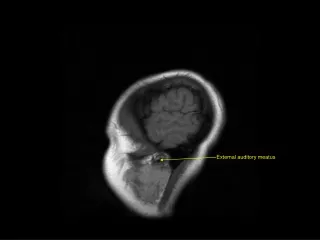
Case Presentation • A 72-year-old male with a 40-year history of recurrent left ear discharge, presented to his private otolaryngologist with left ear discharge, pruritus, and hearing loss. • Examination revealed a firm, granular mass arising from the posterior half of the external auditory canal. • Biopsy of the mass revealed moderately differentiated squamous cell carcinoma. • CT scan and MRI demonstrated soft tissue changes noted in the mastoid and middle ear cleft without temporal bone invasion.
SYMPTOMATOLOGY (Pensak et. Al) • Ear pain 74% • Hearing loss 62% • Bleeding 28% • Facial numbness 12% • Vertigo 10%
Moderately differentiated squamous cell carcinoma in which some, but not all, of the neoplastic cells in nests have pink keratin staining.
CLINICAL SIGNS • Ear canal lesion 88% • Aural discharge 84% • Preauricular swelling 25% • Facial paralysis 18% • Abnormal neck nodes 8%
University of Pittsburgh TNM staging system proposed for the external auditory canal and middle ear. • T1 Tumor limited to the external auditory canal without bony erosion or evidence of soft-tissue extension • T2 Tumor with limited external auditory canal bony erosion (not full thickness) or radiographic finding consistent with limited (<0.5 cm) soft-tissue involvement
University of Pittsburgh TNM staging system proposed for the external auditory canal and middle ear (cont.) • T3 Tumor eroding the osseous external auditory canal (full thickness) with limited (<0.5 cm) soft-tissue involvement, or tumor involving middle ear and/or mastoid, or patients presenting with facial paralysis • T4 Tumor eroding the cochlea, petrous apex, medial wall of middle ear, carotid canal, jugular foramen or dura, or with extensive (>0.5 cm) soft-tissue involvement
University of Pittsburgh TNM staging system proposed for the external auditory canal and middle ear (cont.) • Involvement of lymph nodes is a poor prognostic finding and automatically places the patient in an advanced stage (i.e., Stage III [T1N1] or Stage IV [T2, T3, T4, N1] disease) • Distant metastasis indicates a very poor prognosis and immediately places the patient in the stage IV category.
LITERATURE REVIEW • 1966 - 1996., 96 publications, 144 pts. • No single institution has sufficient data to allow analysis of results. • No randomized or non-randomized prospective studies were identified. • All studies were case series without control subjects.
T E R M I N O L O G Y • MASTOIDECTOMY - all types of modified and radical mastoidectomy. • LATERAL TBR - removal of the osseous and cartilaginous EAC, malleus and incus. • SUBTOTAL TBR - removal of the otic capsule in addition to removal of the osseous and cartilaginous EAC, malleus and incus. • TOTAL TBR - removal of the petrous apex in addition to all that is removed in subtotal resection.
Q U E S T I O N S : 1. What is the survival rate of pts. with EAC, treated by surgical resection and what type of operation should be performed? 2. Once the disease enters the middle ear (ME), what is the operation that provides optimal survival? 3. Is total TBR (Temporal Bone Resection) ever indicated?
Q U E S T I O N S (cont.) : 4. How does prognosis change as structures such as dura mater, brain, and ICA become involved? Is there a role for surgery in these instances? 5. Does the addition of preoperative or postoperative RT enhance survival?
R E S U L T S (1) • E A C - Patients experience similar survival, regardless of whether mastoidectomy, lateral TBR, or subtotal TBR is performed ( 50%, 38.8%, 50% 5-year survival respectively). • The addition of RT to lateral TBR does not appear to improve survival.
R E S U L T S (2) • ME extension - survival of pts. Treated with subtotal TBR (41.7% 5-year survival) appeared to be improved over those treated with lateral TBR (28.6%) or mastoidectomy (17.1%). • Remains uncertain whether the addition of RT to mastoidectomy improves survival.
R E S U L T S (3) • Petrous apex involvement -value of surgical resection remains unclear. subtotal TBR - 0% 1-year survival total TBR - 50% 1-year survival - 0% 2-year survival
R E S U L T S (4) • Resection of involved dura mater does not appear to improve survival. • Incomplete data regarding margins of resection were reported. • Determination of the value of resection of involved brain parenchyma or ICA will require further study.
Testa JR, et al. San Paulo, BrazilArch Otol.Head Neck Surg. 1997 Jul; 123 (7) 720 - 4 79 pts. T1-2 - 34 ; T3-4 - 43 ; Tx - 2; Available - 68 pts. 5 - year survival • Surgery - 65% • Radiotherapy - 29% • Surgery & Radiotherapy - 63%
Hashi N, et al. Hokkaido, Japan. Radiother. Oncol.2000 Aug:56(2) 221-5 20 pts. 8 (T1) - RT alone 65 Gy in 26 fx. Over 6.5 weeks. 12 (T2) - surgery & RT (perioper.) 30 - 75 Gy in 12 - 30 fx. T1 - 100% 5-year survival. T2 - 48% 5-year survival.
Moody SA, et al. Pittsburg, USAAm. J Otol. 2000 Jul; 21(4) 582-8 32 pts. All pts.underwent surgery of the temporal bone and RT (depending on stage and surgical margins) Results: S T A G E 2-YEAR SURVIVAL T1 100% T2 80% T3 50% T4 7%
Perefunder L. et al. Wuerzburg, Germany.Int. J. Radiation Oncology Biol. Phys. Vol.44 No. 4 pp. 777-88, 1999 • 27 pts. • 5-year survival : • T1-2 - 86% T3 - 50% T4 - 41% Complete tumor resection - 100% Positive surgical margins - 66% • 3D CT treatment planing HDR brachytherapy -an effective tool in management of local recurrence following surgery and full course of EBRT.
Radiation technique for treatment of T3N1 carcinoma of the external auditory canal with involvement of an intraparotideal lymph node and perineural tumor spread along the facial nerve.
Dose distribution for brachytherapy treatment of EAC recurrence.Treatment was performed after surgery and a full course of EBRT (70 Gy).
Overall survival of 27 patients with carcinoma of the EAC and middle ear treated at the University of Wuerzburg, and freedom of local recurrence.
Survival of patients with carcinoma of the EAC and middle ear according to T-stages of Pittsburgh classification
Survival of patients with carcinoma of the EAC and middle ear: influence of margins of resection and of intracranial tumor infiltration
C O N C L U S I O N • RT -treatment of choice in T1 disease (Our patient). • Surgery&RT recommended as standard of care for tumors with bony invasion • Survival for T3 tumors is 50% with postoperative RT compared to 0% with surgery alone. (Moody SA et al.) • HDR brachytherapy – is an effective tool in management of local recurrence following surgery and full course EBRT.