Download
1 / 40
410 likes | 613 Views
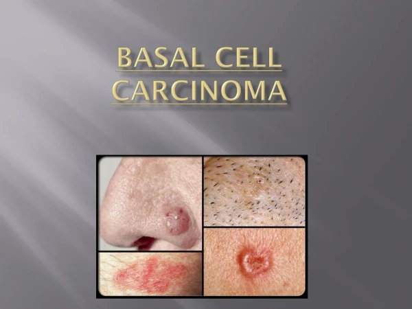
External Ear Basosquamous cell carcinoma. Instructor: 杜宗陽主任 Reporter: 張廷碩. Case Presentation. History. 44-year-old female Sun exposure (+) Left progressive auricular mass for 2 years. 2014.1.14 EAR OPD. Physical Examination.

E N D
External Ear Basosquamous cell carcinoma Instructor: 杜宗陽主任 Reporter: 張廷碩
History • 44-year-old female • Sun exposure (+) • Left progressive auricular mass for 2 years. • 2014.1.14 EAR OPD
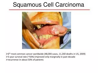
Physical Examination • A PURPLISH MASS LESION ABOUT 2X2 CM OVER LEFT UPPER AURICULAR.
Operation • 2014.1.16: REMOVAL OF EXTERNAL EAR TUMOR +F.T.S.G
Left auricle skin defect • FTSG repair on the skin defect skin
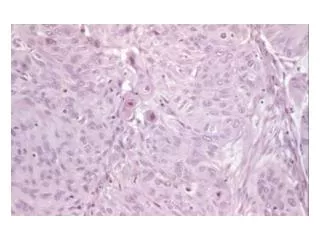
Pathology • It consists of one piece of grey white soft tissue and measures 1.6 x 1.4 x 1.3 cm. • Nodular type basosquamous cell carcinoma. There is no tumor present at the resection margin.
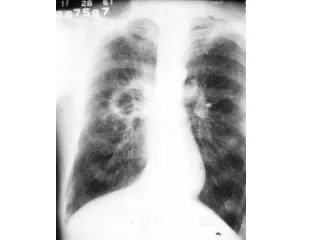
Staging • Neck CT • Abdominal sonogram • WBBS • Staging: pT1cN0M0
Risk Factors • The following are high-risk features for nonmelanoma skin cancer that is not on the eyelid: • The tumor is thicker than 2 millimeters. • The tumor is described as Clark level IV (has spread into the lower layer of the dermis) or Clark level V (has spread into the layer of fat below the skin). • The tumor has grown and spread along nerve pathways. • The tumor began on an ear or on a lip that has hair on it.
Staging • In stage I, The tumor is not larger than 2 centimeters at its widest point and may have one high-risk feature. • In stage II, the tumor is either: • larger than 2 centimeters at its widest point; or • any size and has two or more high-risk features • In stage III: Jaw, eye socket, or side of the skull /LN • In stage IV: base of the skull, spine, or ribs /LN>6cm
Combined Conference • Pathology: • Margin less than 1mm • No perineural invasion • Wider surgical margin is needed. • Post-op R/T is not indicated if margin free.
Operation • 2014.2.6: Tumor wide excision with primary closure
History • First described in 1928. • Today’s definition: WHO 2005 • “Metatypical BCC” : Synonym to Basosquamous Cell Carcinoma • “Keratotic BCC” • “Collision tumor”
Diagnosis • Only made by biopsy. • BCC: • Indolent subtypes: Superficial/Nodular • Aggressive subtypes: Infiltrative/Morpheaform/BSC
Diagnosis • Immunochemical Stain: • Discontinuity of Basement membrane • High percentage of proliferating cells • More stromal reactions
Incidence: 1.2%~2.7% of all skin carcinoma. • Location: head and neck (95%) : Nose (33%) • Increased recurrence and metastatic rates:
Treatment • Moh’s surgery
Treatment • Wide excision with safe margin