Download
1 / 41
500 likes | 1.58k Views
Lumps & Bumps on the Newborn Head. When should I worry?. Joseph A. Garcia-Prats, M.D. Medical Director, Arnold J. Rudolph NICU Professor, Pediatrics & Ethics Baylor College of Medicine. Objectives. Briefly r eview the process of labor Review anatomy of newborn head

E N D
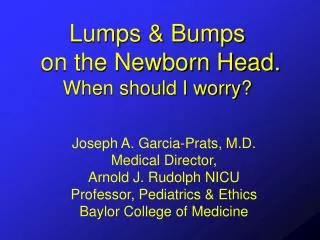
Lumps & Bumps on the Newborn Head.When should I worry? Joseph A. Garcia-Prats, M.D. Medical Director, Arnold J. Rudolph NICU Professor, Pediatrics & Ethics Baylor College of Medicine
Objectives • Briefly review the process of labor • Review anatomy of newborn head • Identify abnormal extra cranial findings • Discuss the most common extra cranial lumps and bumps: Caput succedaneum, cephalohematoma, subgaleal hemorrhage
Process of Labor • Goal of labor is to prepare the cervix and pelvic bones to expel the uterine contents • Describe three phases: latent, active, descent • Approximately 9%- 30% of women have non-vaginal deliveries in the U.S.
Scalp Galea aponeurotica Periosteum Skull bone Dura/pia/arachnoid Brain
Caput Succedaneum Definition: a lesion characterized by a vaguely demarcated area of edema over that portion of the scalp that was the presenting part during a vertex delivery Etiology: extravasation of serum and/or blood from the higher pressures of the uterus and vaginal wall on those areas of the fetal head that border the caput that accumulates over the periosteum
Anatomy of the Scalp Caput succedaneum
Edema Blood Scalp Edema Edema Edema Blood Galea aponeurotica Periosteum Skull bone Dura/pia/arachoid Brain
Caput Succedaneum Occurrence rate: very common Clinical manifestation: soft swelling usually a few millimeters thick (although it may be much thicker) and may be associated with overlying petechiae, purpura or ecchymosis. Extends beyond suture lines.
Caput Succedaneum Treatment: none Resolution: hours to 1-2 days Complications: none
Cephalohematoma Definition: bleeding below the periosteum of the skull Etiology: mechanism of of the bleeding is not exactly known (may occur in uncomplicated vaginal deliveries or in newborns delivered by caesarian section)
Anatomy of the Scalp Cephalo-hematoma
Scalp Galea aponeurotica Periosteum Suture cephalohematoma Suture Skull bone Dura/pia/arachnoid Brain
Cephalohematoma Occurrence: 0.41% - 2.5% of deliveries Noted more often in: (1) males, (2) on right side, (3) newborns delivered vaginally. Bilateral involvement: 15% Diagnosis: “fluid like accumulation” best appreciated at 6-24 hours after delivery; does not transilluminate; boundaries are the suture lines.
Back Front
Cephalohematoma Resolution: 2-8 weeks with a “crater like ridge” noted as it resolves Complications: Hyperbilirubinemia, anemia, infection, calcification, osteomyelitis, skull fracture (5 % occurrence with unilateral and 18% with bilateral cephalohematoma – rarely associated morbidity) Treatment: “Expectant”
Subgaleal Hemorrhage Definition: extracranial bleeding from under the scalp which may become massive and life threatening Etiology: rupture of emissary veins with blood accumulating between the epicranial aponeurosis of the scalp and the periosteum.
Anatomy of the Scalp Subgaleal hemorrhage
Scalp Galea aponeurotica Muscle attach Muscle attach Subgaleal hemorrhage Periosteum Skull bone Dura mater Brain
Subgaleal Hemorrhage Occurrence: RARE -- 1.5 per 10,000 births to 1 per 30,000 births. Appears to be an increased occurrence with vacuum extraction, forceps delivery, but may also be seen in spontaneous deliveries. Contributing factors may be inappropriate placement and/or failed vacuum extraction
Subgaleal Hemorrhage Clinical manifestations: Ill-defined borders,firm to fluctuant, may have fluid waves Potential space includes the limits of: orbital margins back to the nuchal ridge, laterally temporal facia. “Football helmet” like location
Anterior border Posterior border Lateral border
Anterior border Posterior border Lateral border
Subgaleal Hemorrhage Treatment: Close monitoring of vital signs looking for increasing FOC and signs of hypovolemia. Supportive care very important which includes: volume replacement, monitoring for DIC, factor replacement Resolution: 2-3 weeks Complications: Encephalopathy, intracranial pathology(ICH, edema, skull fracture), DIC, jaundice
Anatomy of the Scalp Caput succedaneum Subgaleal hemorrhage Cephalo-hematoma
Edema Edema Blood Scalp Blood Edema Edema Galea aponeurotica Muscle attach Muscle attach Subgaleal hemorrhage Periosteum Suture Suture Cephalohematoma Skull bone Dura mater Brain
In Conclusion • Understanding the anatomy of the tissues surrounding the skull makes it easier to distinguishing the different “lumps and bumps”. • Armed with this knowledge the perinatal health care provider is in a better position to identify patients at risk for complications associated with these lesions.