Download

1 / 48
540 likes | 2.44k Views
NON ACCIDENTAL HEAD INJURY in CHILDREN. Various terms are used – “abusive head trauma, inflicted brain injury, non accidental injury and shaken baby syndrome” Leading cause of infant death from injury Impact and non-impact (shaken). Non Accidental Head injury.

E N D
NON ACCIDENTAL HEAD INJURY in CHILDREN • Various terms are used – “abusive head trauma, inflicted brain injury, non accidental injury and shaken baby syndrome” • Leading cause of infant death from injury • Impact and non-impact (shaken)
Non Accidental Head injury • “The Battered Child Syndrome” Kempe et al 1962 • “The whiplash shaken infant syndrome: manual shaking by the extremities with whiplash induced intracranial and intraocular bleeding, linked with residual permanent brain damage and mental retardation” John Caffey 1974
EPIDEMIOLOGY • Scottish study 25 cases/100,000 children<1 • United Kingdom SDH 21/100,000<1 year • United Kingdom SDH 13/100,000<2 year • North Carolina incidence 17/100,000<2 year with a fatality rate of 22% (3-4/100,000)
NON ACCIDENTAL HEAD INJURY IN CHILDREN; SYDNEY EXPERIENCE • J Neurosurgery (Paediatrics ) 103:213-218, 2005 • Examined the demographics, clinical & radiological features and clinical outcomes of presumed NAI of 65 consecutive cases
Triad of “shaken baby syndrome”John Caffey • Subdural haematomas • Little signs of external trauma • Retinal haemorrhages • Other “red flags” might be changing history and incompatible history – when there is no history of trauma or only minor trauma with subdural haematomas & retinal haemorrhages the PPV for abuse is 0.92
Diagnosis • There is no diagnostic test for non accidental injury • The diagnosis is made on a balance of probability • Careful exclusion of other possible causes eg accidental injury and medical conditions ( rare metabolic disorders, coagulation disorders, infective encephalopathies etc)
Diagnosis • Established by a multidisciplinary team of CPU, neurosurgical, neurology, paediatric, ophthalmology and radiology staff • Detailed family interviews • Exclusion of underlying diseases and accidental injuries
Total = 65 • Range 0.5-46 months • Age 8.2 months (mean) • M:F 39:26
Known in 41/65 cases and usually the male partner (70%) Maternal age 22.6 Male partner 24.5 Risk factors family disruption, separation in 50% cases History of abuse of the child or another family member in 24% Perpetrators
Family Dynamics There was a high incidence of family disruption, substance abuse and prematurity
Seizures 35 patients out of the 65 had seizures on admission or a preceding history before admission and 17 had epilepsy on follow up
Skull Fractures Were seen in 36.9% of cases
Retinal Haemorrhages Seen in 60% of cases and most were bilateral
Retinal Haemorrhages • The subject of controversy. When associated with trauma almost always inflicted injury. But can be associated with increased ICP, coagulopathies, spontaneous SAH, retinal dysplasia, retinopathy of prematurity, galactosaemia, glutaric aciduria and hypertension as well as severe accidental head injury
A History of Trauma Was given in 31 cases (50%) but the mechanism of trauma was inconsistent with the child’s development in the majority of cases (70%) Scalp haematomas 30% Facial bruising 40% Truncal bruising & limb bruising 20%
Presenting symptoms prior to admission for the 65 cases Seizures, irritability, “breathing problems” lethargy, apnoea, sleepy, arrest, poor feeding and failure to thrive, vomiting, a “big head” and coma. # Apnoea (rib fractures) and retinal haemorrhages have a high odds ratio for association with inflicted head injury – a systematic review 2009 (literature review 1970-2008)
Other Injuries • Rib fractures & collapsed lung • Abdominal trauma (hepatic & renal) • Burns to forearm, limbs & trunk • Skeletal injuries > 50% ( often occult)
Skeletal Injuries (60) • > 50% investigated found to have positive bone scans or skeletal survey, although clinical evidence for these injuries existed in only 20% • Multiple areas of skeletal injury in 30% • Limb fractures in 40% • Rib fractures 30% • Facial fractures • Spinal fractures
Admission to Non-accidental Injury • Was made at some stage during the hospitalization in only 14 cases
Hospital Stay • The mean duration of stay was 18 days • Range 1-127 days • Ventilation required in 30 cases (46%)
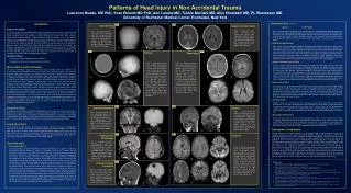
Neuro Imaging • CT Scan - all patients had at least one CT head scan • MRI Scan - 37 patients (57%) also had MRI scans • MRI scanning picked up hypoxic changes, infarction, shearing injury, cerebral contusion & haemorrhage, small subdural haematomas and subarachnoid haemorrhage not seen on CT
MRI Scanning • In summary MRI scans revealed additional pathological findings not visible on CT scanning in 18(49%) of 37 children • Investigation of choice
CT and MRI • Subdural haematoma 53 • Cerebral oedema or ischaemia 22 • Cerebral contusion 21 • Skull fracture 24 • Subarachnoid haemorrhage 12 • Extradural haematoma 1
Characteristics of subdural haematoma • Cerebral convexity 45 • Inter-hemispheric 23 • Posterior fossa 14 • Multiple locations 34 • Acute 10 • Chronic 16 • Differing ages 27
Surgical Procedures • A total of 35 operations were performed in 17 children mainly for subdural and further subdural recollection
Surgical Procedures • Burr-hole drainage of subdural 21 • Insertion of subdural shunt 6 • Insertion of ventricular shunt 2 • External ventricular drain 1 • Removal of shunt 1 • Craniotomy 4
Glasgow Outcome Score 1 Dead 4 2 Vegetative 7 3 Severe Disability 17 4 Moderate Disability 12 5 Good 25
Care Arrangements • After discharge were known for 59 cases and 29 were placed into foster care, 16 to care of relatives and 14 to care of parent
Statistical Analysis Patients were categorized as having a poor outcome if their GOS was 3 or less and a good outcome if 4 or 5 (for statistical analysis) The only statistically significant correlation was seen between CT or MRI findings of ischemia or oedema and outcome
Poor Outcome If there were radiological findings of cerebral ischemia or oedema
Discussion • Risk factors included young parents, unstable family situations, low socioeconomic status and prematurity • The epidemiological characteristics consistent with published data • The most frequent perpetrators were the fathers and the maternal male partners
Discussion • In 24% of cases the families were already known to have a history of abuse • The presentation of NAI or inflicted head injury can be subtle and non specific • An association was seen between the radiological findings of ischemia or oedema and outcome
Discussion • Mortality 6% and severe disability or vegetative 31% • The outcome in this study compares favourably with published data • Recurrence of subdural collections occurs in nearly half of the cases treated with burr-hole drainage
Conclusions Non specific clinical presentation Routine use of MRI to detect ischemia Routine screening of NAI with bone scans High rate of subclinical (occult) skeletal injuries Recurrence of subdural haematomas after surgery Families with history of abuse