Synapses
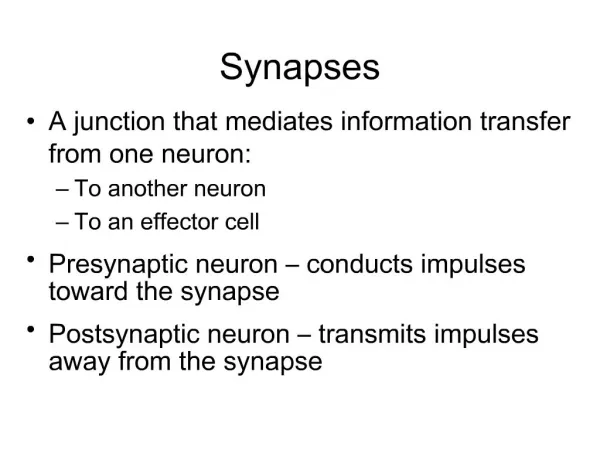
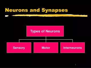
Synapses. A connection that mediates information transfer from one neuron: To another neuron To an effector cell Presynaptic neuron – conducts impulses toward the synapse Postsynaptic neuron – transmits impulses away from the synapse. Synapses. Figure 11.17. Types of Synapses.

Synapses
E N D
Presentation Transcript
Synapses • A connection that mediates information transfer from one neuron: • To another neuron • To an effector cell • Presynaptic neuron – conducts impulses toward the synapse • Postsynaptic neuron – transmits impulses away from the synapse
Synapses Figure 11.17
Types of Synapses • Axodendritic – synapses between the axon of one neuron and the dendrite of another • Axosomatic – synapses between the axon of one neuron and the soma of another • Other types of synapses include: • Axoaxonic (axon to axon) • Dendrodendritic (dendrite to dendrite) • Dendrosomatic (dendrites to soma)
Electrical Synapses • Electrical synapses: • Are less common than chemical synapses • Correspond to gap junctions found in other cell types • Are important in the CNS in: • Arousal from sleep • Mental attention • Emotions and memory • Ion and water homeostasis
Chemical Synapses • Specialized for the release and reception of neurotransmitters • Typically composed of two parts: • Axonal terminal of the presynaptic neuron, which contains synaptic vesicles • Receptor region on the dendrite(s) or soma of the postsynaptic neuron
Synaptic Cleft • Fluid-filled space separating the presynaptic and postsynaptic neurons • Prevents nerve impulses from directly passing from one neuron to the next • Transmission across the synaptic cleft: • Is a chemical event (as opposed to an electrical one) • Ensures unidirectional communication between neurons
Synaptic Cleft: Information Transfer • Nerve impulses reach the axonal terminal of the presynaptic neuron and open Ca2+ channels • Neurotransmitter is released into the synaptic cleft via exocytosis in response to synaptotagmin • Neurotransmitter crosses the synaptic cleft and binds to receptors on the postsynaptic neuron • Postsynaptic membrane permeability changes, causing an excitatory or inhibitory effect
Synaptic Cleft: Information Transfer Figure 11.19
Termination of Neurotransmitter Effects • Neurotransmitter bound to a postsynaptic neuron: • Produces a continuous postsynaptic effect • Blocks reception of additional “messages” • Must be removed from its receptor • Removal of neurotransmitters occurs when they: • Are degraded by enzymes • Are reabsorbed by astrocytes or the presynaptic terminals • Diffuse from the synaptic cleft
Synaptic Delay • Neurotransmitter must be released, diffuse across the synapse, and bind to receptors • Synaptic delay – time needed to do this (0.3-5.0 ms) • Synaptic delay is the rate-limiting step of neural transmission
Postsynaptic Potentials • Neurotransmitter receptors mediate changes in membrane potential according to: • The amount of neurotransmitter released • The amount of time the neurotransmitter is bound to receptors • The two types of postsynaptic potentials are: • EPSP – excitatory postsynaptic potentials • IPSP – inhibitory postsynaptic potentials
Excitatory Postsynaptic Potentials • EPSPs are graded potentials that can initiate an action potential in an axon • Use only chemically gated channels • Na+ and K+ flow in opposite directions at the same time • Postsynaptic membranes do not generate action potentials
Excitatory Postsynaptic Potentials Figure 11.20a
Inhibitory Synapses and IPSPs • Neurotransmitter binding to a receptor at inhibitory synapses: • Causes the membrane to become more permeable to potassium and chloride ions • Leaves the charge on the inner surface negative • Reduces the postsynaptic neuron’s ability to produce an action potential
Inhibitory Synapses and IPSPs Figure 11.20b
Summation • A single EPSP cannot induce an action potential • EPSPs must summate temporally or spatially to induce an action potential • Temporal summation – presynaptic neurons transmit impulses in rapid-fire order
Summation • Spatial summation – postsynaptic neuron is stimulated by a large number of terminals at the same time
Summation Figure 11.21
Neurotransmitters • Chemicals used for neuronal communication with the body and the brain • 50 different neurotransmitters have been identified • Classified chemically and functionally
Chemical Neurotransmitters • Acetylcholine (ACh) • Biogenic amines • Amino acids • Peptides • Novel messengers: ATP and dissolved gases NO and CO
Neurotransmitters: Acetylcholine • First neurotransmitter identified, and best understood • Released at the neuromuscular junction • Synthesized and enclosed in synaptic vesicles • Degraded by the enzyme acetylcholinesterase (AChE) • Released by: • All neurons that stimulate skeletal muscle • Some neurons in the autonomic nervous system
Neurotransmitters: Biogenic Amines • Include: • Catecholamines – dopamine, norepinephrine (NE), and epinephrine • Indolamines – serotonin and histamine • Broadly distributed in the brain • Play roles in emotional behaviors and our biological clock
Synthesis of Catecholamines • Enzymes present in the cell determine length of biosynthetic pathway • Norepinephrine and dopamine are synthesized in axonal terminals • Epinephrine is released by the adrenal medulla Figure 11.22
Neurotransmitters: Amino Acids • Include: • GABA – Gamma ()-aminobutyric acid • Glycine • Aspartate • Glutamate • Found only in the CNS
Neurotransmitters: Peptides • Include: • Substance P – mediator of pain signals • Beta endorphin, dynorphin, and enkephalins • Act as natural opiates, reducing our perception of pain • Bind to the same receptors as opiates and morphine • Gut-brain peptides – somatostatin, and cholecystokinin
Neurotransmitters: Novel Messengers • ATP • Is found in both the CNS and PNS • Produces excitatory or inhibitory responses depending on receptor type • Induces Ca2+ wave propagation in astrocytes • Provokes pain sensation
Neurotransmitters: Novel Messengers • Nitric oxide (NO) • Activates the intracellular receptor guanylyl cyclase • Is involved in learning and memory • Carbon monoxide (CO) is a main regulator of cGMP in the brain
Functional Classification of Neurotransmitters • Two classifications: excitatory and inhibitory • Excitatory neurotransmitters cause depolarizations (e.g., glutamate) • Inhibitory neurotransmitters cause hyperpolarizations (e.g., GABA and glycine)
Functional Classification of Neurotransmitters • Some neurotransmitters have both excitatory and inhibitory effects • Determined by the receptor type of the postsynaptic neuron • Example: acetylcholine • Excitatory at neuromuscular junctions with skeletal muscle • Inhibitory in cardiac muscle
Neurotransmitter Receptor Mechanisms • Direct: neurotransmitters that open ion channels (ligand gated receptors) • Promote rapid responses • Examples: ACh and amino acids • Indirect: neurotransmitters that act through second messengers (metabatropic gated receptors) • Promote long-lasting effects • Examples: biogenic amines, peptides, and dissolved gases
Channel-Linked Receptors • Composed of integral membrane protein • Mediate direct neurotransmitter action • Action is immediate, brief, simple, and highly localized • Ligand binds the receptor, and ions enter the cells • Excitatory receptors depolarize membranes • Inhibitory receptors hyperpolarize membranes
Channel-Linked Receptors Figure 11.23a
G Protein-Linked Receptors: Mechanism Figure 11.23b
G Protein-Linked Receptors: Mechanism • Neurotransmitter binds to G protein-linked receptor • G protein is activated and GTP is hydrolyzed to GDP • The activated G protein complex activates adenylate cyclase • Adenylate cyclase catalyzes the formation of cAMP from ATP • cAMP, a second messenger, brings about various cellular responses
G Protein-Linked Receptors • Responses are indirect, slow, complex, prolonged, and often diffuse • These receptors are transmembrane protein complexes • Examples:neuropeptides receptors, and those that bind biogenic amines
G Protein-Linked Receptors: Effects • G protein-linked receptors activate intracellular second messengers including Ca2+, cGMP, diacylglycerol, as well as cAMP • Second messengers: • Open or close ion channels • Activate kinase enzymes • Phosphorylate channel proteins • Activate genes and induce protein synthesis
Neural Integration: Neuronal Pools • Functional groups of neurons that: • Integrate incoming information • Forward the processed information to its appropriate destination
Neural Integration: Neuronal Pools • Simple neuronal pool • Input fiber – presynaptic fiber • Discharge zone – neurons most closely associated with the incoming fiber • Facilitated zone – neurons farther away from incoming fiber
Types of Circuits in Neuronal Pools • Divergent – one incoming fiber stimulates ever increasing number of fibers, often amplifying circuits Figure 11.25a, b
Types of Circuits in Neuronal Pools • Convergent – opposite of divergent circuits, resulting in either strong stimulation or inhibition Figure 11.25c, d
Types of Circuits in Neuronal Pools • Reverberating – chain of neurons containing collateral synapses with previous neurons in the chain Figure 11.25e
Types of Circuits in Neuronal Pools • Parallel after-discharge – incoming neurons stimulate several neurons in parallel arrays Figure 11.25f
Patterns of Neural Processing • Serial Processing • Input travels along one pathway to a specific destination • Works in an all-or-none manner • Example: spinal reflexes
Patterns of Neural Processing • Parallel Processing • Input travels along several pathways • Pathways are integrated in different CNS systems • One stimulus promotes numerous responses • Example: a smell may remind one of the odor and associated experiences
Development of Neurons • The nervous system originates from the neural tube and neural crest • The neural tube becomes the CNS • There is a three-phase process of differentiation: • Proliferation of cells needed for development • Migration – cells become amitotic and move externally • Differentiation into neuroblasts
Axonal Growth • Guided by: • Scaffold laid down by older neurons • Orienting glial fibers • Release of nerve growth factor by astrocytes • Neurotropins released by other neurons • Repulsion guiding molecules • Attractants released by target cells
N-CAMs • N-CAM – nerve cell adhesion molecule • Important in establishing neural pathways • Without N-CAM, neural function is impaired • Found in the membrane of the growth cone