Download
1 / 34
340 likes | 439 Views
Learn about different types of muscles such as skeletal, cardiac, and smooth, their functions, anatomy at gross and microscopic levels, muscle contraction mechanisms, neuromuscular junction, muscle metabolism, fiber types, and interactions of skeletal muscles.

E N D
Muscle Types • Skeletal: striated, voluntary • Cardiac: only in heart, striated, involuntary • Smooth/Visceral: walls of organs, not striated, involuntary
Functions • Movement • Posture maintenance • Heat generation (3/4 of energy produced by ATP escapes as heat) • Stabilization of joints • Protection of some internal organs
Skeletal Muscle Gross Anatomy
Muscle: Organ • Consists of hundreds to thousands of muscle cells (fibers) • Covered by epimysium (connective tissue that binds muscles into functional groups) • Blood vessels and nerve fibers • Fascicle: portion of muscle (bundle of muscle cells surrounded by perimysium)
Skeletal Muscle Fiber = Muscle Cell • Striated, elongated, multinucleate • Surrounded by endomysium (connective tissue) or sarcolemma • Sarcoplasmic reticulum (SR) inside each muscle cell: set of interconnecting tubules • Composed of actin & myosin
Microscopic Anatomy • Myofibril: complex organelle composed of bundles of myofilaments; banded • Sarcomere: contractile unit composed of myofilaments made of contractile protein
Myofilaments: Two Types • Actin (thin) filament: long bead like strands (twisted double strand of pearls); tropomyosin and troponin on beaded strand • Myosin (thick) filament: rod-like tail with two globular heads
A Bands • 1 sarcomere • Extends from Z line to next Z line • Contains both actin and myosin
I Bands • Contain actin
Contraction of Muscle Fiber • Sarcomeres shorten myofibrils shorten • Muscle Mechanics Skit from STARS program
Sliding Filament Theory of Contraction • Crossbridge Attachment: activated myosin heads are strongly attracted to exposed binding sites on actin and crossbridge binding occurs • Power Stroke: as myosin head binds, it changes from high energy configuration to its bent, low-energy shape, which causes head to pull on thin filament, sliding it toward center of sarcomere
As new ATP molecule binds to myosin head, myosin crossbridge is released from actin • Cocking of Myosin Head: hydrolysis of ATP to ADP and Pi provides energy needed to return myosin head to its high energy or cocked position, which gives it potential energy needed for next attachment
Neuromuscular Junction • Site where nerve & muscle fiber meet • Acetylcholine (ACh): neurotransmitter that relays message from nerve to muscle fiber • Acetylcholinesterase (AChE): An enzyme that breaks down ACh
Regulation of Contraction Mechanism • ACh is released at neuromuscular junction • Calcium diffuses • Tropomyosin moves and exposes active sites on actin • Linkages form between actin and myosin • Muscle fiber shortens • AChE is released and decomposes ACh • Muscle fiber relaxes
Motor Unit • Motor neuron and all the muscle fibers it supplies • Fine control: fingers, eyes < 150 muscle fibers per motor neuron • Less precise control: hips, legs > 150 muscle fibers per motor neuron
Graded Muscle Responses • Variations in degree (strength & length) of muscle contraction • Requirement for proper control of skeletal movement
Muscle Metabolism • Energy for contraction: ATP, glucose, and glycogen • Muscle fatigue: glucose from blood and reserve glycogen are exhausted; lactic acid buildup; ATP cannot keep pace
Slow-Twitch Fatigue Resistant Fibers • Red color reflects plentiful supply of myoglobin which stores oxygen • Abundant mitochondria • Good blood supply • Specialized for endurance • Example: long distance runners
Fast Twitch Fatigable Fibers • White fibers • Contract rapidly • Few mitochondria but large glycogen reserves • Extremely powerful but fatigue quickly • Example: sprinters
Fast Twitch Fatigue Resistant Fibers • In between slow twitch and fast twitch fatigable in power and endurance
Microscopic Structure & Arrangement of Smooth Muscle • Spindle shaped cells • One centrally located nucleus • No striations present
Types of Smooth Muscles • Varies in the following ways: • Fiber arrangement and organization • Responsiveness to various stimuli • Innervation
Single-Unit Smooth Muscle • More common • Also called visceral muscle • Contract as unit and rhythmically “talk” to one another through gap junctions • Arranged in sheets
Multiunit Smooth Muscle • Located in large airways of lungs, arrector pili, internal eye (pupil) and large arteries • No gap junctions so they act as individual cells • Many nerve ending attachments
Interactions of Skeletal Muscles • Groups of muscles work either together or in opposition to achieve a wide variety of movements • Muscles can only pull, NEVER push • Contraction ONLY • Insertion: attachment on movable bone • Origin: fixed or immovable point of attachment
Prime Movers/Agonists: assume major responsibility for movement • Antagonists: muscles that oppose, or reverse, a particular movement • Synergists: promote the same movement or reduce undesirable movements • Fixators: immobilize a bone or muscle’s origin
Movements at Joints • Flexion: decreases angle between two bones • Extension: increases angle between two bones • Hyperextension: increases angle between two bones beyond anatomical position • Dorsiflexion: moves the sole of the foot upward • Plantar flexion (extension): moves the sole of the foot downward as in standing on the toes
Adduction: moving a body part toward the midline • Abduction: moving a body part away from the midline • Circumduction: the distal end of an extremity inscribes a circle while the shaft inscribes a cone
Rotation: revolving a part about the longitudinal axis • Supination: turn the palm upward • Pronation: turn the palm downward • Inversion: turn the plantar surface away from the midline
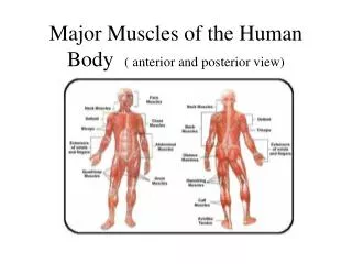
Major Muscles of Movement • Gluteus maximus • Gluteus medius • Gluteus minimus