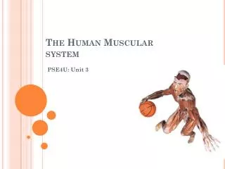
MUSCULAR SYSTEM: Histology and Physiology
MUSCULAR SYSTEM: Histology and Physiology. Introduction. Types and Features of Muscle Tissue smooth muscle : cardiac muscle : muscle :. skeletal. General Characteristics of Muscle contractility excitability. extensibility. elasticity. General Functions of Muscle. 1.


MUSCULAR SYSTEM: Histology and Physiology
E N D
Presentation Transcript
Introduction • Types and Features of Muscle Tissue • smooth muscle: • cardiac muscle: • muscle: skeletal
General Characteristics of Muscle • contractility • excitability extensibility elasticity
General Functions of Muscle • 1. • 2. • 3. • 4.
Skeletal Muscle: Structure organs • Skeletal muscles are composed of skeletal muscle fibers, connective tissue, blood vessels and nerves.
Muscle fiber , is a skeletal muscle cell • single, cylindrically shaped cell • multiple nuclei, located peripherally • development • develop from • myoblasts fuse to form muscle cells • grow into skeletal muscle to form neuromuscular junction • One neuron per skeletal muscle fiber • numbers of striated muscle cells remains relatively constant after birth, but they may enlarge myoblasts Motor nerves
size of muscle fibers/cells • 1 - 40 mm long • 10 - 100 microns in diameter • size of fibers varies with size of muscle • muscle fibers are (banded appearance) striated
sarcolemma • Muscle Anatomy • - plasma membrane of muscle cell
endomysium • also a delicate layer of CT with reticular fibers • surrounds each muscle fiber outside the external lamina • CT layer • surrounds a bundle of muscle fibers, a perimysium muscle fasciculus
epimysium fasiculi • a relatively thick layer of dense irreg. collagenous CT • surrounds the many that make up a muscle • covers the entire surface of the muscle
epimysium • fascia • a layer of fibrous CT outside the • separates individual muscles • may surround muscle groups • fascia of an individual muscle is also called the epimysium
these CT layers are continuous with each other and with the tendons and CT sheaths of the bones • functions of CT • holds muscles together • provides a passageway to the muscle cells for blood vessels and nerves • tendon or sheetlike aponeurosis • epimysium fused to periosteum indirect attachment direct attachment
Muscle Fibers, Myofibrils, & Myofilaments • Muscle cell components • sarcolemma sarcoplasm
myofibrils • threadlike structures, 1 - 3 micrometers in diameter • extend from one end of the muscle fiber to another • composed of protein myofilaments • - thin filaments • - thick filaments actin myosin
Z line to Z • sarcomere • a highly organized unit of myofilaments • join end to end with other sarcomeres to form myofibrils • extends from line (line=disc) • Movie
I (isotropic) band • banding due to arrangements of actin & myosin microfilaments • light band • includes a Z lines (disk) • consists only of actin filaments • extends on either side of Z line to myosin myofilaments
A (anisotropic) band • dark band • extends the length of myosin myofilaments within the sarcomere
H zone • a smaller band located within the center of each A-band • only myosin myofilaments are present, no overlapping with actin
(desmin) • M line • a dark band in the middle of the H zone • consists of delicate filaments that attach to the center of the myosin myofilaments • holds myosin myofilaments in place
titin (elastic filament) • an elastic filament anchoring myosin myofilament at M line to Z disc • resists excessive stretching or sarcomere
Actin myofilaments • two strands of fibrous actin (F-actin) for double helix • which are polymers of about 200 globular ( ) monomers G-actin
active site • two strands are arranged in a double helix • each G-actin monomer has an to which myosin molecules can bind during muscle contraction
tropomyosin • molecules • an elongated molecule that winds along the groove of the F-actin double helix • each molecule covers 7 G-actin active sites
troponin • composed of three subunits • one with a high affinity for actin (TnI) • one with a high affinity for tropomyosin (TnT) • one with a high affinity for ions (TnC) • spaced between ends of tropomyosin molecules in groove between F-actin double helix calcium
in presence of Ca2+, troponin is activated • troponin activation results in it displacing tropomyosin deeper into the “groove” in F-actin double helix • this exposes the actin active sites
Myosin myofilaments • composed of elongated, club-shaped, myosin molecules • two parts of myosin molecule • rods (tails) • with ATPase heads
rod-like portions wound together • about 100 myosin molecules per myosin myofilament • point of attachment between head and rod is hinged • myosin head forms cross bridge (contact) to actin’s active site
striated pattern • other • A bands and I bands of parallel myofibrils are aligned to produce seen with a microscope • many • many glycogen granules & lipid droplets mitochondria
transverse (T) tubules • tube like invaginations of the sarcolemma • project into the muscle fiber and wrap around the sarcomeres where the actin and myosin microfilaments overlap • lumen is continuous with exterior of muscle fiber • filled with fluid extracellular
sarcoplasmic reticulum • highly specialized smooth ER • near the T tubules, enlarged to form terminal cisternae • is a T tubule and 2 terminal cisternae • transports ions from sarcoplasm to its lumen triad calcium
Clinical Focus • Muscular dystrophy • Muscular atrophy Robbins
Sliding Filament Theory • Contracting Muscle • actin and myosin myofilaments don't change length but slide past one another during muscle contraction. • cross bridges form between heads of myosin and active sites on actin, release, and then reform, causing the actin myofilaments at each end of the sarcomere to slide past the myosin microfilaments toward the H zone • I bands and H zones become more narrow during contraction; H zone may disappear • A bands remain constant in length • sarcomere shortens
Slidng filament theory 1 • Slidng filament theory 2
Neuromuscular Junctions • Motor neurons • specialized nerve cells that propagate action potentials to skeletal muscle fibers at a relatively high velocity • most motor neurons enter skeletal muscles with the blood vessels branching when they reach the • each motor neuron innervates more than muscle fiber • a branch of the motor neuron innervates one muscle fiber, forming a neuromuscular junction or synapse near the center of the muscle fiber perimysium single
Neuromuscular junction structure • presynaptic (axonal) terminal • postsynaptic terminal (motor end plate) • synaptic cleft • synaptic vesicles: Release acetylcholine
Quick Review • Membrane Transport 2 • Active processes • Move substances “uphill” or against their concentration gradient; most often is the energizer for this process • Active Transport is the mechanism • ; an mechanism ATP Na+ - K+ ATPase antiport
Na+ - K+ ATPase 1 • Na+ - K+ ATPase 2
Resting Membrane Potential • The cell membrane is more permeable to some molecules than to others. • Membrane Potentials • A , or electrical potential due to the separation of charges by the cell membrane. • Resting Membrane Potential occur in all cells due to • RMP ranges from -20 to -200 millivolts (mV) • state of the cell Voltage Concentration Gradients Active Transport Pumps Electrical Gradients Polarized
Inside of the cell is electrically neutral • Outside of the cell is electrically neutral • But, • higher outside of cell • Membrane ~impermeable to Na+ • higher inside of cell • Membrane ~permeable to K+ • K+ diffuses down its concentration gradient • This concentration difference make a loss of positive charges inside the cell • maintains this “imbalance” How? [Na+] [K+] Na+ - K+ ATPase
Electric charges • But there is more, • reinforce the ion concentration on the inside and outside of the cell • Electrical forces resist K+ diffusion out of the cell • An produces the RMP electrochemical gradient Fig. 9.7
Action Potential tracing • Stimulus • Na+ channel open -> Na+ influx • K+ channel begin to open late in phase • Na+ channel close • K+ channel open • (after potential) • K+ leaks until Na+-K+ ATPase reestablish RMP Depolarization phase Repolarization phase Hyperpolarization
Regulation - Skeletal Muscle Fiber • Regulation of Contraction • Nerve Stimulus & Neuromuscular Junction • has cell body in CNS (brain & spinal cord) Motor neuron