Download
1 / 24
240 likes | 385 Views
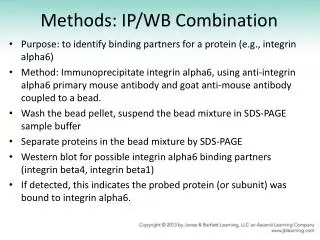
Methods: IP/WB Combination. Purpose: to identify binding partners for a protein (e.g., integrin alpha6) Method: Immunoprecipitate integrin alpha6, using anti-integrin alpha6 primary mouse antibody and goat anti-mouse antibody coupled to a bead.

E N D
Methods: IP/WB Combination • Purpose: to identify binding partners for a protein (e.g., integrin alpha6) • Method: Immunoprecipitate integrin alpha6, using anti-integrin alpha6 primary mouse antibody and goat anti-mouse antibody coupled to a bead. • Wash the bead pellet, suspend the bead mixture in SDS-PAGE sample buffer • Separate proteins in the bead mixture by SDS-PAGE • Western blot for possible integrin alpha6 binding partners (integrin beta4, integrin beta1) • If detected, this indicates the probed protein (or subunit) was bound to integrin alpha6.
iClicker time The (approximate) wavelengths of visible light are shown in the table at left. Based on these values, what do you feel would be the best filter pair for visualizing Green Fluorescent Protein? A. 400 nm excitation filter, 700 nm emission filter B. 540 nm excitation filter, 570 nm emission filter C. 450 nm excitation filter, 530 nm emission filter D. 625 excitation filter, 540 nm emission filter E. 700 excitation filter, 500 nm emission filter
Logical Argument • A formal construction for reaching logical conclusions • An argument consists of two parts: • At least TWO premises (statements of fact) • A conclusion Premise A Premise B, etc. Conclusion
Logical Argument • In science, the premises are supplied by data • Be careful about over-interpreting your data (an image is just an image, not a functional test…) • In research articles, premises are supplied mostly by figures in the article. • Summarizing the data in the figures supplies the premises for the overall argument of a study • Arguments can be nested, meaning the conclusion of one argument can be a premise for the next argument. • Goal for Exam 1: Understand the logic of Figure 1 in the paper by Carien M. Niessen et al.
Chapter 4 Phospholipids and Membrane Structure George Plopper
Learning Outcomes • Define a hypothesis • Provide an example of a logical argument • Draw a phospholipid containing one cis-unsaturated fatty acid and one saturated fatty acid • Diagram the current fluid mosaic model of a membrane • Define the role of the endoplasmic reticulum in membrane synthesis
Hypothesis • In science, a hypothesis is NOT a prediction; instead, it is a proposed explanation for an observed set of facts • What’s the difference? If a prediction is wrong, nobody learns anything; if an explanation is wrong, it helps clarify the problem to increase the likelihood of finding the correct explanation.
Figure 04.05: Phospholipid bilayers have varying permeability to solutes.
Figure 04.07: Three classes of membrane proteins. Each class is represented by a different colored protein.
Figure 04.08A: The orientation of alpha helices and beta sheets in transmembrane portions of integral membrane proteins.
Figure 04.08B: The orientation of alpha helices and beta sheets in transmembrane portions of integral membrane proteins.
Figure 04.09: Plasma membrane proteins form clusters that permit cell adhesion.
Figure 04.10: GPI is synthesized and attached to membrane proteins in the ER.
Figure 04.11: Three mechanisms for translocation of phospholipids from one leaflet of the ER membrane to the other.
Figure 04.12: Establishment and maintenance of lipid asymmetry in the plasma membrane.
Figure 04.13: Protein "tags" direct some cytosolic proteins to enter organelles.
Figure 04.14: Sterol carrier protein-2 structure. Note the fatty acid bound to it.
Figure 04.15: Summary of membrane synthesis mechanisms in eukaryotes.
Table 04.T01: Relative composition of phospholipids in representative membranes.