Download
1 / 97
980 likes | 1.33k Views
Chapter 42. Circulation and Gas Exchange. Overview: Trading Places. Every organism must exchange materials with its environment. Exchanges ultimately occur at the cellular level. In unicellular organisms, these exchanges occur directly with the environment.

E N D
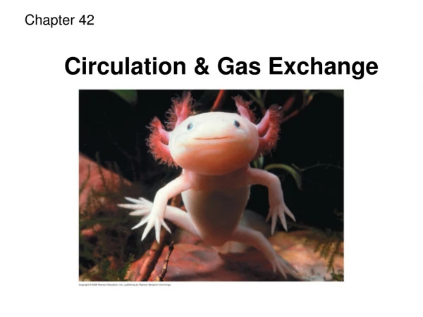
Chapter 42 Circulation and Gas Exchange
Overview: Trading Places • Every organism must exchange materials with its environment. • Exchanges ultimately occur at the cellular level. • In unicellular organisms, these exchanges occur directly with the environment.
For most cells making up multicellular organisms, direct exchange with the environment is not possible. • Gills are an example of a specialized exchange system in animals. • Internal transportandgas exchangeare functionally related in most animals.
Circulatory systems link exchange surfaces with cells throughout the body • In small and/or thin animals, cells can exchange materials directly with the surrounding medium. • In most animals, transport systems connect the organs of exchange with the body cells. • Most complex animals have internal transport systems that circulate fluid.
Gastrovascular Cavities • Simple animals, such as cnidarians, have a body wall that is only two cells thick and that encloses a gastrovascularcavity. • This cavity functions in both digestionanddistribution of substances throughout the body. • Some cnidarians, such as jellies, have elaborate gastrovascular cavities. • Flatworms have a gastrovascular cavity and a large surface area to volume ratio.
Internal transport in gastrovascular cavities Circular canal Mouth Pharynx Mouth Radial canal 5 cm 2 mm (a) The moon jelly Aurelia, a cnidarian (b) The planarian Dugesia, a flatworm
Open and Closed Circulatory Systems • More complex animals have either open or closed circulatory systems. • Both systems have three basic components: • A circulatory fluid = blood or hemolymph. • A set of tubes = blood vessels. • A muscular pump = the heart.
In insects, other arthropods, and most molluscs, blood bathes the organs directly in an open circulatory system. • In an open circulatory system, there is no distinction between blood and interstitial fluid, and this general body fluid is more correctly called hemolymph.
In a closed circulatory system, the blood is confined to vessels and is distinct from the interstitial fluid. • Closed systems are more efficient at transporting circulatory fluids to tissues and cells.
Open and closed circulatory systems Heart Heart Blood Hemolymph in sinuses surrounding organs Small branch vessels In each organ Interstitial fluid Pores Dorsal vessel (main heart) Tubular heart Auxiliary hearts Ventral vessels (a) An open circulatory system (b) A closed circulatory system
Organization of Vertebrate Closed Circulatory Systems • Humans and other vertebrates have a closed circulatory system, often called the cardiovascular system. • The three main types of blood vessels are: arteries - away from the heart. veins- toward the heart. capillaries - exchange with body cells.
Arteries branch into arterioles and carry blood to capillaries. • Networks of capillaries called capillary bedsare the sites of chemical exchange betweenthebloodandinterstitial fluid. • Venules converge into veins and return blood from capillaries to the heart.
Vertebrate hearts contain two or more chambers. • Blood enters through an atrium and is pumped out through a ventricle. Atria-receive blood Ventricles-pump blood
Single Circulation • Bony fishes, rays, and sharks have single circulation with a two-chambered heart. • In single circulation, blood leaving the heart passes through two capillary beds before returning.
Single circulation in fishes Gill capillaries Gill circulation Artery Ventricle Heart Atrium Systemic circulation Vein Systemic capillaries
Double Circulation • Amphibian, reptiles, and mammals have double circulation. • Oxygen-poor and oxygen-rich blood are pumped separately from the right and left sides of the heart.
Double circulation in vertebrates Mammals and Birds Amphibians Reptiles Lung and skin capillaries Lung capillaries Lung capillaries Right systemic aorta Pulmocutaneous circuit Pulmonary circuit Pulmonary circuit Atrium (A) Atrium (A) A A A A V V Ventricle (V) V V Left systemic aorta Left Right Left Right Right Left Systemic circuit Systemic circuit Systemic capillaries Systemic capillaries Systemic capillaries
In reptiles and mammals, oxygen-poor blood flows through the pulmonary circuitto pick up oxygen through the lungs. • In amphibians, oxygen-poor blood flows through a pulmocutaneous circuit to pick up oxygen through the lungs and skin. • Oxygen-rich blood delivers oxygen through the systemic circuit. • Double circulation maintains higher blood pressure in the organs than does single circulation.
Adaptations of Double Circulatory Systems Amphibians: • Frogs / amphibians have a three-chambered heart: 2 atria and 1 ventricle. • The ventricle pumps blood into a forked artery that splits the ventricle’s output into the pulmocutaneous circuit and the systemic circuit. • Underwater, blood flow to the lungs is nearly shut off.
Reptiles (Except Birds) • Turtles, snakes, and lizards have a three-chambered heart: two atria and one ventricle. • In alligators, caimans, and other crocodilians a septum - partially or fully divides the ventricle. • Reptiles have double circulation, with a pulmonary circuit - lungs and a systemic circuit.
Mammals RA --> RV --> LUNGS --> LA --> LV --> Body • Mammals and birds have a four-chambered heart with two atria and two ventricles. • The left side of the heart pumps and receives only oxygen-rich blood, while the right side receives and pumps only oxygen-poor blood. • Mammalsandbirds are endothermsand require more O2 than ectotherms.
Coordinated cycles of heart contraction drive double circulation in mammals • Blood begins its flow with the right ventricle pumping blood to the lungs. • In the lungs, the blood loads O2 and unloads CO2 • Oxygen-rich blood from the lungs enters the heart at the left atrium and is pumped through the aorta to the body tissues by the left ventricle. • The aorta provides blood to the heart through the coronary arteries.
Blood returns to the heart through the superior vena cava (deoxygenated blood from head, neck, and forelimbs) and inferior vena cava (deoxygenated blood from trunk and hind limbs). • The superior vena cava and inferior vena cava flow into the Right Atrium - RA.
Capillaries of head and Forelimbs - EXCHANGE Superiorvena cava Returns deoxygenated blood from body to heart RA 7 Pulmonary artery Carries deoxygenated blood to lungs Pulmonary artery Capillaries of right Lung GAS EXCHANGE Aorta 9 Capillaries of left Lung GAS EXCHANGE 3 3 2 4 11 Pulmonary vein Carries oxygenated blood to heart: LA Pulmonary vein 5 1 Right Atrium RA - Receives deoxygenated blood from body Left Atrium - LA Receives oxygenated blood from lungs 10 Right Ventricle RV - Pumps blood to lungs Left Ventricle - LV Pumps oxygenated blood to body Inferior vena cava Returns deoxygenated blood from body to heart RA Aorta = main artery to body for Systemic Circulation mammalian cardiovascular system Capillaries of abdominal organs and hind limbs EXCHANGE with body cells 8
The Mammalian Heart: A Closer Look • A closer look at the mammalian heart provides a better understanding of double circulation. • RIGHT side= deoxygenated blood from body pumped to lungs. • LUNGS = gas exchange. • LEFT side = oxygenated blood from lungs pumped to body.
Mammalian Heart Aorta -systemic circulation Pulmonary artery - to lungs Right Atrium RA Receives Deoxygented Blood from body Pulmonary veins - from lungs to heart Left Atrium LA Receives oxgenated blood from lungs Semilunar valve Semilunar valve Atrioventricular valve Atrioventricular valve Right Ventricle RV Pumps to lungs for gas exchange Left Ventricle LV Pumps oxygenated blood to body via aorta
The heart contracts and relaxes in a rhythmic cycle called the cardiac cycle. • The contraction, or pumping, phase is called systole. • The relaxation, or filling, phase is called diastole. • Blood Pressure = systolic / diastolic
Cardiac cycle Atrial systole; ventricular diastole 2 Semilunar valves closed 0.1 sec Semilunar valves open AV valves open 0.4 sec 0.3 sec 1 Atrial and ventricular diastole AV valves closed 3 Ventricular systole; atrial diastole
The heart rate, also called the pulse, is the number of beats per minute. • The stroke volumeis the amount of blood pumped in a single contraction. • The cardiac outputis the volume of blood pumped into the systemic circulationper minute and depends on both the heart rate and stroke volume.
Four valves prevent backflow of blood in the heart: • The atrioventricular (AV) valves separate each atrium and ventricle. • The semilunar valves control blood flow to the aorta and the pulmonary artery. • The “lub-dup” sound of a heart beat is caused by the recoil of blood against the AV valves (lub) then against the semilunar (dup) valves. • Backflow of blood through a defective valve causes a heart murmur.
Maintaining the Heart’s Rhythmic Beat • Some cardiac muscle cells are self-excitable = they contract without any signal from the nervous system. • The sinoatrial (SA) node, or pacemaker, sets the rate and timing at which cardiac muscle cells contract. • Impulses from the SA node travel to the atrioventricular (AV) node. At the AV node, the impulses are delayed and then travel to the Purkinje fibers that make the ventricles contract. • Impulses that travel during the cardiac cycle can be recorded as an electrocardiogram(ECG or EKG). The pacemaker is influenced by nerves, hormones, body temperature, and exercise.
Control of heart rhythm 3 1 2 Pacemaker generates wave of signals to contract. Signals are delayed at AV node. Signals pass to heart apex. Signals spread throughout ventricles. 4 AV node SA node (pacemaker) Bundle branches Purkinje Fibers: ventricles contract Heart apex ECG
Patterns of blood pressure and flow reflect the structure and arrangement of blood vessels • The physical principles that govern movement of water in plumbing systems also influence the functioning of animal circulatory systems. • The epithelial layer that lines blood vessels is called the endothelium.
Structure of blood vessels Artery Vein SEM Valve 100 µm Basal lamina Endothelium Endothelium Smooth muscle Smooth muscle Connective tissue Connective tissue Capillary Artery Vein Arteriole Venule 15 µm Red blood cell Capillary LM
Capillaries have thin walls, the endothelium plus its basement membrane, to facilitate the exchange of materials. • Arteries and veins have an endothelium, smooth muscle, and connective tissue. • Arteries have thicker walls than veins to accommodate the high pressure of blood pumped from the heart. • In the thinner-walled veins, blood flows back to the heart mainly as a result of muscle action.
Blood Flow Velocity • Physical laws governing movement of fluids through pipes affect blood flow and blood pressure. • Velocity of blood flow is slowest in the capillary beds,as a result of thehigh resistanceand large total cross-sectional area. • Blood flow in capillaries is necessarily slow for exchange of materials.
The interrelationship of cross-sectional area of blood vessels, blood flow velocity, and blood pressure. 5,000 4,000 Area (cm2) 3,000 2,000 1,000 0 50 40 Velocity (cm/sec) 30 20 10 0 120 Systolic pressure 100 80 Pressure (mm Hg) 60 Diastolic pressure 40 20 0 Aorta Veins Arteries Venules Arterioles Capillaries Venae cavae
Blood Pressure • Blood pressure is the hydrostatic pressure that blood exerts againstthe wallof avessel. • In rigid vessels blood pressure is maintained; less rigid vessels deform and blood pressure is lost.
Changes in Blood Pressure During the Cardiac Cycle • Systolic pressureis the pressure in the arteries during ventricle contraction /systole; it is the highest pressure in the arteries. • Diastolic pressureis the pressure in the arteries during relaxation /diastole; it is lower than systolic pressure. • A pulseis the rhythmic bulging of artery walls with each heartbeat.
Regulation of Blood Pressure • Blood pressure is determined by cardiac output and peripheral resistance due to constriction of arterioles. • Vasoconstrictionis the contraction of smooth muscle in arteriole walls; it increases blood pressure. • Vasodilationis the relaxation of smooth muscles in the arterioles; it causes blood pressure to fall.
Vasoconstriction and vasodilation help maintain adequate blood flow as the body’s demands change. • The peptide endothelin is an important inducer of vasoconstriction. • Blood pressure is generally measured for an artery in the arm at the same height as the heart. • Blood pressure for a healthy 20 year old at rest is 120mm Hg at systole / 70 mm Hg at diastole.
Question: How do endothelial cells control vasoconstriction? RESULTS Ser Leu Endothelin Ser Met Cys —NH3+ Ser Cys Asp Lys Glu Ile —COO– Cys Val Tyr Phe Cys His Leu Asp Ile Trp Cys Trp Parent polypeptide 53 73 1 203 Endothelin
Measurement of blood pressure: sphygmomanometer Blood pressure reading: 120/70 Pressure in cuff greater than 120 mm Hg Pressure in cuff drops below 120 mm Hg Pressure in cuff below 70 mm Hg Rubber cuff inflated with air 120 120 70 Artery closed Sounds audible in stethoscope Sounds stop
Fainting is caused by inadequate blood flow to the head. • Animals with longer necks require a higher systolic pressure to pump blood a greater distance against gravity. • Blood is moved through veins by smooth muscle contraction, skeletal muscle contraction, and expansion of the vena cava with inhalation. • One-way valves in veins / heart prevent backflow of blood.
Blood flow in veins Direction of blood flow in vein (toward heart) Valve (open) Skeletal muscle Valve (closed)
Capillary Function • Capillaries in major organs are usually filled to capacity. Blood supply varies in many other sites. • Two mechanisms regulate distribution of blood in capillary beds: • Contraction of the smooth muscle layer in the wall of an arteriole constricts the vessel. • Precapillary sphincters control flow of blood between arterioles and venules.
Blood flow in capillary beds Thoroughfare channel Precapillary sphincters Capillaries Arteriole Venule (a) Sphincters relaxed Arteriole Venule (b) Sphincters contracted
The critical exchangeof substances betweenthe blood andinterstitial fluid takes place across the thin endothelial walls of the capillaries. • The difference between blood pressureand osmotic pressure drives fluids out of capillaries at the arteriole end and into capillaries at the venule end.
Fluid exchange between capillaries and the interstitial fluid Body tissue INTERSTITIAL FLUID Capillary Net fluid movement out Net fluid movement in Direction of blood flow Blood pressure = hydrostatic pressure Inward flow Pressure Outward flow Osmotic pressure Arterial end of capillary Venous end