Circulation and Gas Exchange Overview: Organisms' Environmental Trading
Explore how organisms exchange materials with their environment at the cellular level in this detailed overview of circulation systems and gas exchange processes.


Circulation and Gas Exchange Overview: Organisms' Environmental Trading
E N D
Presentation Transcript
Chapter 42 Circulation and Gas Exchange
Overview: Trading with the Environment • Every organism must exchange materials with its environment • And this exchange ultimately occurs at the cellular level
In unicellular organisms • These exchanges occur directly with the environment • For most of the cells making up multicellular organisms • Direct exchange with the environment is not possible
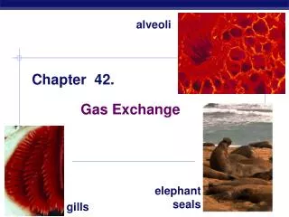
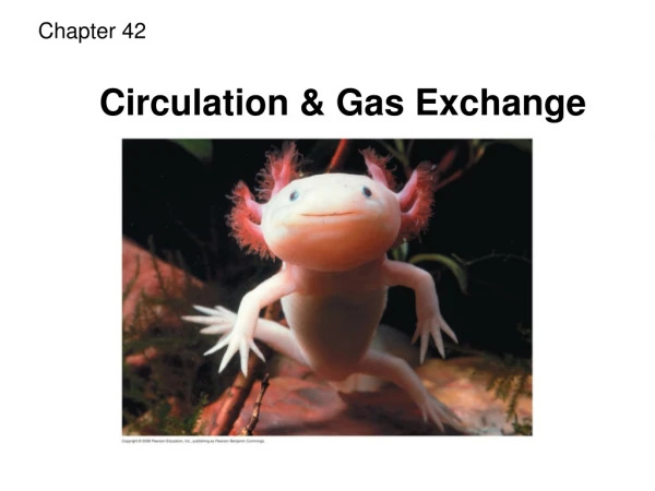
Figure 42.1 • The feathery gills projecting from a salmon • Are an example of a specialized exchange system found in animals
Concept 42.1: Circulatory systems reflect phylogeny • Transport systems • Functionally connect the organs of exchange with the body cells
Most complex animals have internal transport systems • That circulate fluid, providing a lifeline between the aqueous environment of living cells and the exchange organs, such as lungs, that exchange chemicals with the outside environment
Invertebrate Circulation • The wide range of invertebrate body size and form • Is paralleled by a great diversity in circulatory systems
Gastrovascular Cavities • Simple animals, such as cnidarians • Have a body wall only two cells thick that encloses a gastrovascular cavity • The gastrovascular cavity • Functions in both digestion and distribution of substances throughout the body
Circularcanal Radial canal Mouth 5 cm Figure 42.2 • Some cnidarians, such as jellies • Have elaborate gastrovascular cavities
Open and Closed Circulatory Systems • More complex animals • Have one of two types of circulatory systems: open or closed • Both of these types of systems have three basic components • A circulatory fluid (blood) • A set of tubes (blood vessels) • A muscular pump (the heart)
Heart Hemolymph in sinusessurrounding ograns Ostia Anterior vessel Lateral vessels Tubular heart Figure 42.3a (a) An open circulatory system • In insects, other arthropods, and most molluscs • Blood bathes the organs directly in an open circulatory system
Heart Interstitialfluid Small branch vessels in each organ Dorsal vessel(main heart) Ventral vessels Auxiliary hearts (b) A closed circulatory system Figure 42.3b • In a closed circulatory system • Blood is confined to vessels and is distinct from the interstitial fluid
Closed systems • Are more efficient at transporting circulatory fluids to tissues and cells
Survey of Vertebrate Circulation • Humans and other vertebrates have a closed circulatory system • Often called the cardiovascular system • Blood flows in a closed cardiovascular system • Consisting of blood vessels and a two- to four-chambered heart
Arteries carry blood to capillaries • The sites of chemical exchange between the blood and interstitial fluid • Veins • Return blood from capillaries to the heart
Fishes • A fish heart has two main chambers • One ventricle and one atrium • Blood pumped from the ventricle • Travels to the gills, where it picks up O2 and disposes of CO2
Amphibians • Frogs and other amphibians • Have a three-chambered heart, with two atria and one ventricle • The ventricle pumps blood into a forked artery • That splits the ventricle’s output into the pulmocutaneous circuit and the systemic circuit
Reptiles (Except Birds) • Reptiles have double circulation • With a pulmonary circuit (lungs) and a systemic circuit • Turtles, snakes, and lizards • Have a three-chambered heart
Mammals and Birds • In all mammals and birds • The ventricle is completely divided into separate right and left chambers • The left side of the heart pumps and receives only oxygen-rich blood • While the right side receives and pumps only oxygen-poor blood
A powerful four-chambered heart • Was an essential adaptation of the endothermic way of life characteristic of mammals and birds
Gill capillaries Lung and skin capillaries Lung capillaries Lung capillaries AMPHIBIANS REPTILES (EXCEPT BIRDS) MAMMALS AND BIRDS FISHES Right systemicaorta Pulmonarycircuit Artery Pulmocutaneouscircuit Pulmonarycircuit Gillcirculation Heart:ventricle (V) Left Systemicaorta A A A A A A Atrium (A) V V V V V Left Right Left Left Right Right Systemiccirculation Systemic circuit Systemic circuit Vein Systemic capillaries Systemic capillaries Systemic capillaries Systemic capillaries Figure 42.4 • Vertebrate circulatory systems
Concept 42.2: Double circulation in mammals depends on the anatomy and pumping cycle of the heart • The structure and function of the human circulatory system • Can serve as a model for exploring mammalian circulation in general
Mammalian Circulation: The Pathway • Heart valves • Dictate a one-way flow of blood through the heart
Blood begins its flow • With the right ventricle pumping blood to the lungs • In the lungs • The blood loads O2 and unloads CO2
Oxygen-rich blood from the lungs • Enters the heart at the left atrium and is pumped to the body tissues by the left ventricle • Blood returns to the heart • Through the right atrium
Capillaries of head and forelimbs Anterior vena cava Aorta Pulmonary artery Pulmonary artery 9 6 3 3 7 8 Capillaries of right lung Capillaries of left lung 2 4 11 Pulmonary vein Pulmonary vein Left atrium 5 1 Right atrium 10 Left ventricle Right ventricle Aorta Posterior vena cava Capillaries of abdominal organs and hind limbs Figure 42.5 • The mammalian cardiovascular system
Pulmonary artery Aorta Pulmonaryveins Pulmonaryartery Anterior vena cava Leftatrium Right atrium Pulmonaryveins Atrioventricularvalve Semilunarvalve Semilunarvalve Atrioventricularvalve Posterior vena cava Right ventricle Figure 42.6 Left ventricle The Mammalian Heart: A Closer Look • A closer look at the mammalian heart • Provides a better understanding of how double circulation works
The heart contracts and relaxes • In a rhythmic cycle called the cardiac cycle • The contraction, or pumping, phase of the cycle • Is called systole • The relaxation, or filling, phase of the cycle • Is called diastole
Atrial systole; ventricular diastole 2 Semilunarvalvesclosed 0.1 sec Semilunarvalvesopen 0.3 sec 0.4 sec AV valvesopen Atrial and ventricular diastole AV valvesclosed 1 3 Ventricular systole; atrial diastole Figure 42.7 • The cardiac cycle
The heart rate, also called the pulse • Is the number of beats per minute • The cardiac output • Is the volume of blood pumped into the systemic circulation per minute
Maintaining the Heart’s Rhythmic Beat • Some cardiac muscle cells are self-excitable • Meaning they contract without any signal from the nervous system
A region of the heart called the sinoatrial (SA) node, or pacemaker • Sets the rate and timing at which all cardiac muscle cells contract • Impulses from the SA node • Travel to the atrioventricular (AV) node • At the AV node, the impulses are delayed • And then travel to the Purkinje fibers that make the ventricles contract
The impulses that travel during the cardiac cycle • Can be recorded as an electrocardiogram (ECG or EKG)
Signals pass to heart apex. Signals spread Throughoutventricles. Pacemaker generates wave of signals to contract. Signals are delayed at AV node. Bundlebranches AV node SA node(pacemaker) Purkinjefibers Heartapex ECG 2 1 3 4 Figure 42.8 • The control of heart rhythm
The pacemaker is influenced by • Nerves, hormones, body temperature, and exercise
Concept 42.3: Physical principles govern blood circulation • The same physical principles that govern the movement of water in plumbing systems • Also influence the functioning of animal circulatory systems
Blood Vessel Structure and Function • The “infrastructure” of the circulatory system • Is its network of blood vessels
Vein Artery Valve Endothelium Basementmembrane Smoothmuscle 100 µm Endothelium Connectivetissue Endothelium Smoothmuscle Capillary Connectivetissue Artery Vein Venule Arteriole Figure 42.9 • All blood vessels • Are built of similar tissues • Have three similar layers
Structural differences in arteries, veins, and capillaries • Correlate with their different functions • Arteries have thicker walls • To accommodate the high pressure of blood pumped from the heart
Direction of blood flowin vein (toward heart) Valve (open) Skeletal muscle Valve (closed) Figure 42.10 • In the thinner-walled veins • Blood flows back to the heart mainly as a result of muscle action
Blood Flow Velocity • Physical laws governing the movement of fluids through pipes • Influence blood flow and blood pressure
5,0004,000 3,000 2,000 1,000 0 Area (cm2) 5040 30 20 10 0 Velocity (cm/sec) 120100 80 60 40 20 0 Systolicpressure Pressure (mm Hg) Diastolicpressure Aorta Veins Arteries Venules Capillaries Arterioles Venae cavae Figure 42.11 • The velocity of blood flow varies in the circulatory system • And is slowest in the capillary beds as a result of the high resistance and large total cross-sectional area
Blood Pressure • Blood pressure • Is the hydrostatic pressure that blood exerts against the wall of a vessel
Systolic pressure • Is the pressure in the arteries during ventricular systole • Is the highest pressure in the arteries • Diastolic pressure • Is the pressure in the arteries during diastole • Is lower than systolic pressure
1 3 A typical blood pressure reading for a 20-year-oldis 120/70. The units for these numbers are mm of mercury (Hg); a blood pressure of 120 is a force that can support a column of mercury 120 mm high. 4 The cuff is loosened further until the blood flows freely through the artery and the sounds below the cuff disappear. The pressure at this point is the diastolic pressure remaining in the artery when the heart is relaxed. Blood pressure reading: 120/70 Pressurein cuff above 120 Pressurein cuff below 120 Pressurein cuff below 70 Rubber cuffinflatedwith air 120 120 70 Sounds stop Sounds audible instethoscope Artery Arteryclosed A stethoscope is used to listen for sounds of blood flow below the cuff. If the artery is closed, there is no pulse below the cuff. The cuff is gradually deflated until blood begins to flow into the forearm, and sounds from blood pulsing into the artery below the cuff can be heard with the stethoscope. This occurs when the blood pressure is greater than the pressure exerted by the cuff. The pressure at this point is the systolic pressure. A sphygmomanometer, an inflatable cuff attached to apressure gauge, measures blood pressure in an artery.The cuff is wrapped around the upper arm and inflated until the pressure closes the artery, so that no blood flows past the cuff. When this occurs, the pressure exerted by the cuff exceeds the pressure in the artery. 2 Figure 42.12 • Blood pressure • Can be easily measured in humans
Blood pressure is determined partly by cardiac output • And partly by peripheral resistance due to variable constriction of the arterioles
Capillary Function • Capillaries in major organs are usually filled to capacity • But in many other sites, the blood supply varies
Two mechanisms • Regulate the distribution of blood in capillary beds • In one mechanism • Contraction of the smooth muscle layer in the wall of an arteriole constricts the vessel
Thoroughfare channel Precapillary sphincters (a) Sphincters relaxed Venule Arteriole Capillaries (b) Sphincters contracted Venule Arteriole (c) Capillaries and larger vessels (SEM) Figure 42.13 a–c 20 m • In a second mechanism • Precapillary sphincters control the flow of blood between arterioles and venules
The critical exchange of substances between the blood and interstitial fluid • Takes place across the thin endothelial walls of the capillaries