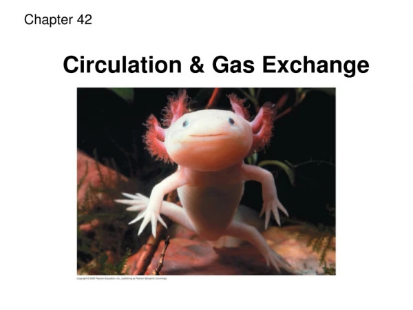
Chapter 42
Chapter 42. Urinalysis. Urine Formation. Urine forms in kidneys and leaves body through urethra The urinary system >>. Urine Formation. How body excretes water and gets rid of waste Waste can become toxic if not removed Two kidneys eliminate soluble waste products of metabolism

Chapter 42
E N D
Presentation Transcript
Chapter 42 Urinalysis
Urine Formation • Urine forms in kidneys and leaves body through urethra The urinary system >>
Urine Formation • How body excretes water and gets rid of waste • Waste can become toxic if not removed • Two kidneys eliminate soluble waste products of metabolism • Click here for an animation
Urine Formation • Filtration • Glomerulus filters waste products, salts, and excess fluid from blood • Tubule concentrates filtered material • Nephron • Combination of glomerulus and tubule • One million nephrons in each kidney
Urine Formation • Filtration • Substances filtered out from body • Water • Ammonia • Electrolytes • Glucose
Urine Formation • Filtration • Substances filtered out from body • Amino acids • Creatinine • Urea • Diabetes diagnosis • Routine urinalysis testing
Urine Formation • Reabsorption • About 180 liters of filtrate produced daily • Only 1–2 liters of urine eliminated • Much filtrate reabsorbed into body • Blood cells and most proteins stay in blood • Concentration of glucose in blood below 180 mg/dl will be reabsorbed • Glucose is a threshold substance
Urine Formation • Secretion • Substances not already filtered are secreted into urine through distal convoluted tubule • Hydrogen and ammonium ions may be secreted into urine in exchange of sodium
Urine Composition • After passing through kidney, urine is 96% water and 4% dissolved substances (urea, salt, sulfates, phosphates)
Urine Composition • Abnormal constituents of urine • WBC • Fat • Glucose • Casts • Bile • Hemoglobin and RBC
Urine Composition • Changes in urine production • Amount of urine excreted can rise or fall • Urine color can change • Urine appearance can vary
Urine Composition • Changes in urine production • Urine odor can change • Cells can be present in urine • Chemical constituents in urine can change • Urine concentration (specific gravity) may vary
Safety • Standard precautions • Transmission-based precautions • Biohazard precautions • Proper disposal of urine
Quality Control • Regulatory agencies • Written testing protocols • Maintained testing records • Recalibration of instruments
Quality Control • Documentation of daily control testing must be kept at least 3 years • Commercially available urine control samples • Run positive and negative controls each day on all tests
CLIA 88 • Appropriate training in methodology of test being performed • Understanding of urine testing quality control procedures • Proficiency in the use of instrumentation; being able to troubleshoot problems
CLIA 88 • Knowledge of stability and proper storage of reagents • Awareness of factors that influence test results • Knowledge of how to verify test results • CLIA categorizes microscopic exam as a PPMP
Urine Containers • Types • Nonsterile containers for cultures • 24 hour collection containers with added preservatives
Urine Containers • Label container immediately after specimen collection • Patient’s name, age, gender, identifying number • Date and time of collection • Physician’s name • Label the cup, not the lid
Urine Collection • Giving patient instructions
Urine Collection • Click to play the video
Urine Collection • Urine specimen types • Random (spot) specimen • Obtained at any time • Most common • If concentrated specimen preferred, first specimen of day is most concentrated
Urine Collection • Urine specimen types • Fasting/timed specimens • Used when physician wants to measure substance without interference from food intake • Length of fast varies • Give patient written directions • Use regular urinalysis container
Urine Collection • Urine specimen types • 24-hour specimen • Circadian rhythm and intake of food and water determine concentration of substances at different times during day/night • Requested when quantitative tests for different substances are desired • Expressed in units per 24 hours • Use of preservatives and refrigeration • Sometimes use 2-hour or 12-hour collection instead
Urine Collection • Urine specimen types • Catheterized specimen • Insert sterile tube directly into bladder through urethra • Not contaminated • Can cause infection if not done correctly • Use only when other methods are contraindicated or show repeated positive testing for bacteria
Urine Collection • Collection methods • Random collection • Clean-catch method; midstream collection • Catheterized
Examination of Urine • Best when fresh, even still warm • Test within 30 minutes, or refrigerate
Routine Urinalysis Procedure • Physical examination of urine • Assess volume of urine specimen, making sure specimen is sufficient for testing • Note any unusual urine odor • Measure specific gravity of specimen
Routine Urinalysis Procedure • Physical examination of urine • Observe and record color and transparency of specimen
Measuring Specific Gravity • Urinometer • Measures specific gravity • Reading the meniscus • Take temperature of urine into account
Measuring Specific Gravity • Refractometer • Most common tool for measuring specific gravity of liquids • Measures refractive index of urine • Reads about 0.002 below that of true specific gravity • Needs 1 drop of urine • Easy to use but more expensive
Routine Urinalysis Procedure • Chemical examination of urine • Use of multistix reagent strips with color-coded chart • Chemical testing available on urine reagent test strips • See Table 42-3
Routine Urinalysis Procedure • Chemical examination of urine • Reagent test strip quality control • Automated urine analyzers >>
Routine Urinalysis Procedure • Microscopic examination of urine sediment • Classified as PPMP • Sediment is forced to the bottom of centrifuged tube • Helps determine kidney disease, disorders of urinary tract, and systemic disease • Need fresh urine • Use of urine color atlas • Use of urine stains
Preparing for Microscopic Examination • Centrifuge 10–15 mL of urine • Pour off supernatant urine • Resuspend sediment by tapping • Stain (optional) >> • Put drop of sediment on slide
RBC WBC Renal epithelial cells Bacteria Yeast Parasites Sperm Artifacts Squamous epithelial cells Urine Sediment Cells and Microorganisms
Crystals in Urine Sediment • Require little attention • Form as urine specimens stand • Uric acid, cystine, and sulfa drug crystals can indicate disease states
Casts in Urine Sediment • Important to note • Formed when protein accumulates and precipitates in kidney tubules • Appearance of casts • Hyaline cast most common kind seen • Granular casts and cellular casts also seen • Takes an experienced eye to identify
Urinalysis Report • Include patient’s name, type of specimen, collection method, ordering provider, MA name, date and time of collection, date and time of testing, findings
Drug Screening • Becoming more common for employment • Test itself is CLIA waived, but detailed protocols must be followed • Chain of custody