Ascending Sensory Pathways
Ascending Sensory Pathways. Dorsal Column-Medial Lemniscus system fine touch position sense Anterolateral system temperature coarse touch pain. James Bisley (jbisley@mednet.ucla.edu). Dorsal Column-Medial Lemniscus system.

Ascending Sensory Pathways
E N D
Presentation Transcript
Ascending Sensory Pathways Dorsal Column-Medial Lemniscus system fine touch position sense Anterolateral system temperature coarse touch pain James Bisley (jbisley@mednet.ucla.edu)
Dorsal Column-Medial Lemniscus system Conveys mechanosensory information from the periphery to the cortex • Cutaneous Mechanoreceptors (fine touch) • Proprioception & Kinesthesia (position)
Pain Temperature Coarse touch receptor afferent Fine touch
Position sense Kinesthesia is the “awareness” of body position and movement Proprioception is the “sub-concious” information used in the feed-back control of posture and precise movements.
Position sense Position sense information comes from: Muscle spindles Golgi tendon organs Joint receptors Cutaneous mechanoreceptive afferents Efference copy
Proprioceptors Motor unit (controlled by efferent) Muscle spindle Golgi tendon organ Joint receptor
First order neuron Second order neuron Third order neuron receptor Dorsal Column-Medial Lemniscus system Some terminology We use the terms first, second and third order neurons to describe the steps of the pathway to cortex.
Dorsal Column-Medial Lemniscus system Afferents have their cell bodies in the DORSAL ROOT GANGLIA. Called pseudo-unipolar neurons.
Spinal reflexes, Clarke’s Nucleus, etc Dorsal Column-Medial Lemniscus system The DRG axons enter through the dorsal horn of the spinal cord
Dorsal Column-Medial Lemniscus system Fibers that convey information from lower limbs and body (below spinal segment T6) travel ipsilaterally along the GRACILE TRACT.
GRACILE TRACT Dorsal Column-Medial Lemniscus system Fibers that convey information from upper limbs and body (above spinal segment T6) travel ipsilaterally along the CUNEATE TRACT. There is a topographic representation of the body in the dorsal columns
Dorsal Column-Medial Lemniscus system Fibers in the Gracile Tract have their first synapse in the GRACILE NUCLEUS. Fibers in the Cuneate Tract have their first synapse in the CUNEATE NUCLEUS. There is a topographic representation of the body in the dorsal column nuclei Caudal medulla
Dorsal Column-Medial Lemniscus system Axons from the second order neurons form the INTERNAL ARCUATE FIBERS in the caudal medulla, which decussates becoming the contralateral MEDIAL LEMNISCUS. There is a topographic representation of the body in the medial lemniscus Caudal medulla
Dorsal Column-Medial Lemniscus system The representation of the body shifts as the medial lemniscus runs rostrally.
Dorsal Column-Medial Lemniscus system The axons of the second order neurons terminate in the VENTRAL POSTERIOR LATERAL NUCLEUS of the thalamus (VPL). There is a topographic representation of the body in the VPL (lower extremities are lateral)
Cuneate Gracile Dorsal Column-Medial Lemniscus system What about the face?
Dorsal Column-Medial Lemniscus system What about the face? Pseudo-unipolar neurons have their cell bodies in the TRIGEMINAL GANGLION. Except for Proprioception Pseudo-unipolar neurons have their cell bodies in the MESENCEPHALIC NUCLEUS inside the CNS. Mid-pons
Dorsal Column-Medial Lemniscus system What about the face? Pseudo-unipolar neurons have their cell bodies in the TRIGEMINAL GANGLION. Except for Proprioception Pseudo-unipolar neurons have their cell bodies in the MESENCEPHALIC NUCLEUS inside the CNS.
Dorsal Column-Medial Lemniscus system What about the face? Axons project to second order neurons in the PRINCIPAL (SENSORY) NUCLEUS OF THE TRIGEMINAL COMPLEX in mid-pons. Mid-pons There is a topographic representation of the face in the principal (sensory) nucleus
Dorsal Column-Medial Lemniscus system What about the face? Axons of the second order neurons decussate and join the TRIGEMINOTHALAMIC TRACT (which runs adjacent to the medial lemniscus). Mid-pons
Dorsal Column-Medial Lemniscus system What about the face? The axons of the second order neurons terminate in the VENTRAL POSTERIOR MEDIAL NUCLEUS of the thalamus (VPM). Mid-pons There is a topographic representation of the face in the VPM
Dorsal Column-Medial Lemniscus system Neurons in the VP complex project to PRIMARY SOMATIC-SENSORY CORTEX via the POSTERIOR LIMB of the INTERNAL CAPSULE. Mid-pons The whole body is represented in the ventral posterior complex.
Dorsal Column-Medial Lemniscus system Neurons in the VP complex project to PRIMARY SOMATIC-SENSORY CORTEX via the POSTERIOR LIMB of the INTERNAL CAPSULE.
Dorsal Column-Medial Lemniscus system Area 3a Primarily proprioception input Area 3b Primarily tactile input Area 1 Primarily tactile input, but receptive fields usually cover several digits Area 2 Combination of tactile and proprioception. Hand configuration & stimulus shape are both important
Dorsal Column-Medial Lemniscus system The whole body is represented in each area of SI Owl Monkey
Dorsal Column-Medial Lemniscus system The somatosensory homunculus
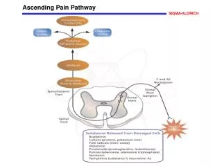
Anterolateral system Conveys pain, temperature and coarse touch information from the periphery to the cortex
Central Pain Pathways: Sensory discriminative component As with the tactile system, the cell bodies are located in the DORSAL ROOT GANGLIA. Pseudo-unipolar neurons.
Central Pain Pathways: Sensory discriminative component The DRG axons enter through the dorsal horn of the spinal cord Upon entering, the axons branch into ascending and decending collaterals forming the DORSOLATERAL TRACT of LISSAUER.
Central Pain Pathways: Sensory discriminative component The axons run up or down several spinal cord segments in Lassauer’s tract before synapsing in the gray matter of the dorsal horn.
anterior white commissure Central Pain Pathways: Sensory discriminative component The second order neurons decussate immediately and form the SPINOTHALAMIC TRACT (aka the anterolateral tract).
anterior white commissure Central Pain Pathways: Sensory discriminative component The second order neurons decussate immediately and form the SPINOTHALAMIC TRACT (aka the anterolateral tract). There is a topographic representation of the body in the spinothalamic tract
Central Pain Pathways: Sensory discriminative component The second order neurons decussate immediately and form the SPINOTHALAMIC TRACT (aka the anterolateral tract).
Central Pain Pathways: Sensory discriminative component The second order neurons decussate immediately and form the SPINOTHALAMIC TRACT (aka the anterolateral tract).
Central Pain Pathways: Sensory discriminative component The second order neurons decussate immediately and form the SPINOTHALAMIC TRACT (aka the anterolateral tract).
Central Pain Pathways: Sensory discriminative component The second order neurons decussate immediately and form the SPINOTHALAMIC TRACT (aka the anterolateral tract).
Just like the tactile system Central Pain Pathways: Sensory discriminative component Neurons in the spinothalamic tract terminate in the VENTRAL POSTERIOR LATERAL NUCLEUS (VPL) of the Thalamus. There is a topographic representation of the body in the VPL (lower extremities are lateral)
Dorsal column Anterolateral Some simple differences between the pathways X X Test the pathway Light touch Vibration 2-point discrimination Sense of position Test the pathway Pain Temperature Coarse touch
Pseudo-unipolar neurons have their cell bodies in the TRIGEMINAL GANGLIONand ganglia associated with nerves VII (Facial), IX (Glosso-pharyngeal) & X (Vagus). Anterolateral tract Central Pain Pathways: Sensory discriminative component What about the face?
Central Pain Pathways: Sensory discriminative component After entering the brain stem, the fibers descendin the SPINAL TRIGEMINAL TRACT to the medulla, where they synapse onto neurons in the SPINAL NUCLEUS of the TRIGEMINAL COMPLEX (primarily the pars caudalis). Anterolateral tract There is a topographic representation of the head in the pars caudalis
Central Pain Pathways: Sensory discriminative component Axons from the second order neurons decussate immediately and then join the ascending anterolateral tract in the brain stem.
Central Pain Pathways: Sensory discriminative component Axons from the second order neurons terminate in the VENTRAL POSTERIOR MEDIAL NUCLEUS (VPM) of the Thalamus. Anterolateral tract There is a topographic representation of the face in the VPM
Central Pain Pathways: Sensory discriminative component The whole body & all somatic senses are represented in the ventral posterior complex. Neurons in the VP complex carrying pain information project to PRIMARYandSECONDARYSOMATIC-SENSORY CORTEX.
Central Pain Pathways: Sensory discriminative component Cortex localization of pain Sub-cortical perception of pain Paleospinothalamic pathways suffering component of pain (reduced by benzodiazepines)
Central Pain Pathways: Descending Control of Pain The same holds true for the pars caudalis of the spinal nucleus of the trigeminal complex Stimulation of PAG results in analgesia.
Endogenous opioid Central Pain Pathways: Descending Control of Pain In the dorsal horn or the pars caudalis Opioids play a role in the descending control of pain
Central Pain Pathways: Local Control of Pain Interaction between dorsal column and anterolateral systems regulates pain perception. This is why rubbing a wound after sharp pain helps a bit. Cutaneous mechanoreceptor Cutaneous nociceptor Stimulation of dorsal columns can antidromically induce analgesia
Central Pain Pathways: Local & Descending Control of Pain What you should know Aα and Aβ fibers excite interneurons that reduce the transmission of pain information Descending fibers excite interneurons that reduce the transmission of pain information
Dorsal Column-Medial Lemniscus system • Information content • Fine touch, vibration and sense of position • The path to cortex • Locations & projections of 1st, 2nd & 3rd order neurons • Where decussation occurs • Differences between DRG inputs & Vth nerve inputs • Basic arrangement of topography throughout the system • The organization of somatosensory cortex • 4 areas • Basic arrangement of topography