Download
1 / 54
591 likes | 1.07k Views
Musculoskeletal System. Sugito Wonodirekso , MS, Dr Department of Histology FMUI. Materials. Skeletal muscle Joint Joint types Bone Cartilages Supporting tissues. Objectives of the muscle tissue. Identify skillfully the skeletal muscle structure

E N D
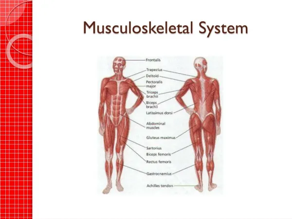
Musculoskeletal System SugitoWonodirekso, MS, Dr Department of Histology FMUI
Materials • Skeletal muscle • Joint • Joint types • Bone • Cartilages • Supporting tissues Musculoskeletal System
Objectives of the muscle tissue • Identify skillfully the skeletal muscle structure • Identify the structural and functional different between 3 major types of muscle tissue • Comprehend the relationships between muscle fascicles, muscle fibers, myofibrils, and myofilaments • Explain the structure and function of T-tubule in skeletal muscle • Analyze the relationships between normal structure and function of skeletal muscle • Explain the regeneration process of skeletal muscle Musculoskeletal System
General features of muscle tissues • Terminology • Prefixes: Sarco- and or myo- • Specialized for contraction • Myofilaments: actin (thin) and myosin (thick) • Mesodermal origin • Exception: iris smooth muscle arise from ectoderm • Cell shape • May reach 4 cm long called fibers (myofibers) • Organization • Works in groups or separately • Two major types • Smooth and striated Musculoskeletal System
Muscle types and characteristics Musculoskeletal System
Skeletal muscle (this is our concern now) • Histogenesis • Mesenchymal cells of mesodermal origin fuse to each other to make • Myoblasts which then fuse to make • Myotubes which later • Elongate by incorporating additional myoblasts • Eventually accumulated myofilaments which are organized into myofibrils and displaced nuclei and other cytoplasmic components peripherally Musculoskeletal System
Skeletal muscle cells • Mature skeletal muscle fibers: • Elongated • Unbranched • Cylindrical • Multinucleated • Flattened peripherally displaced nuclei, lie just under sarcolemma (muscle cell plasma membrane) • Most organelles and sarcoplasm (muscle cells cytoplasm) are displaced near the nuclei’s poles • Sarcoplasm contains mitochondria, glycogen granules, and myoglobin (oxygen-binding protein). It accumulates lipofuscin pigment with age • Mature skeletal cell are end cells and cannot divide Musculoskeletal System
Skeletal muscle tissue • Cross-cut of skeletal muscle to show connective tissue partitioning of muscle into groups or bundles of fibers. Endomysium is very delicate and lies between individual fibers, while perimysium is more visible and lies around a group of fibers. Epimysium is not seen here but ensheaths a whole muscle. In this picture notice the presence of small blood vessels in both perimysium and endomysium. Notice also the cross-cuts of myofibrils within the muscle cells, making them look grainy. Musculoskeletal System
Higher power of skeletal muscle for details of cross-striations. Notice thin Z discs and heavy A bands. From one Z disc to the next is a sarcomere, the unit of muscle contraction. In the upper muscle cell notice shadowy myofibrils running longitudinally. Musculoskeletal System
Skeletal muscle cells (fibers), with cross-striations and peripheral nuclei. Musculoskeletal System
Muscle fibers organization Musculoskeletal System
Sarcomeres (contraction units) Musculoskeletal System
Sarcomere and the cross sections Musculoskeletal System
Myofilament • Thin filaments (actin) • Filamentous actin (F-actin) is polymeric chain of globular actin(G-actin)monomer. Each thin filament consist of 2 double helix wound F-actin strands • Tropomyosinis long, thin, double-helical polipeptides that wrap around the actin double helix, lies in grooves on its surface, and spans 7 G-actin monomers • Troponin is a complex of 3 globular proteins. • TnT (Troponin T) attaches each complex to specific site on each tropomyosin molecule, • TnC binds calcium ions, and • TnI inhibits the interaction between the thin and thick filaments Musculoskeletal System
Actin filaments Musculoskeletal System
Myofilament • Thick filaments (myosin): • Long golf-club-shaped polypeptide • A bundles of myosin molecules with their shafts pointing toward and overlapping in the bundle’s middle and their heads projecting from the bundle’s ends • This arrangement leaves a headless region in the center of each filament corresponding to the H band • Treating myosin molecule with papain (a proteolytic enzyme) cleaves them, at a point near head, into 2 pieces • The piece containing most of the thin shaft is termed light meromyosin; the head and the associated portion of the shaft make up the heavy meromyosin • The head portion of heavy meromyosin has an ATP-binding site and an actin binding site, which are necessary for contraction Musculoskeletal System
Actin and myosin filaments relationship Musculoskeletal System
Myofilament • Organization • The banding pattern of skeletal muscle reflects the grouping of myofilaments into parallel bundles of thin and thick filaments called myofibrils. Each muscle fiber may contain several myofibrils; the number depending on its size. • Take special attention on the appearance of myofibrils in cross- and longitudinal section, especially in EM images and its schematic version Musculoskeletal System
Sarcomere and muscle contraction • Diagram of contraction of skeletal muscle. On the left is the view with light microscopy. On the right are the thin actin filaments and thick myosin filaments seen in EM. Notice that the total width of the A band stays the same throughout and that the sliding in or out of the actin filaments determines the width of the H band. Consider which filaments you would see if you cut the muscle cross-wise through the I band, A band, or H band. Musculoskeletal System
T-tubules and the Triads • Drawing of relationship (at EM level) of myofibrils to sarcoplasmic reticulum (smooth ER) and T-tubules in skeletal muscle. In this drawing the sarcoplasmic reticulum is labelled "sarcotubules" and "terminal cisternae". Notice that T-tubules are extensions of the sarcolemma (cell membrane, seen at right-hand edge), so that depolarization can spread along this part of the sarcolemma as well. (See diagrams and further explanation in your textbook.) Musculoskeletal System
The sarcomere and the diads • Same diagram, for cardiac muscle. • Note differences with skeletal muscle in: • their amount and arrangement of sarcoplasmic reticulum • the presence or near-absence of terminal cisterns (next to the T-tubules) • the position of T-tubules in relation to the A, I, and Z bands seen at the left. • A triad consists of two terminal cisterns with a T-tubule in the middle. When the cisterns are not well developed, a true triad does not exist. A diad means two elements are together, as with one T-tubule and a neighboring bit of sarcoplasmic reticulum. NOTE: sarcoplasmic reticulum is just a form of smooth endoplasmic reticulum (SER). In muscle it is particularly associated with the release of calcium ions needed for contraction. Musculoskeletal System
The sarcomere • EM of several myofibrils running longitudinally through skeletal muscle cell. Between individual myofibrils lie the mitochondria (M) and glycogen (G) of the cytoplasm. Within each myofibril are the typical striations: • A= A band; • I= I band; • Z= Z line; and • H= H band. • The banding is formed by the arrangement of myosin and actin filaments. Musculoskeletal System
Sarcomere and the contraction Musculoskeletal System
Skeletal muscle regeneration Musculoskeletal System
Skeletal muscle regeneration Musculoskeletal System
Skeletal muscle regeneration Musculoskeletal System
Contraction process-1 Musculoskeletal System
Contraction process-2 Musculoskeletal System
Contraction process-3 Musculoskeletal System
Muscle fibers organization Musculoskeletal System
Joints Basic joint components are: Bone Hyaline Cartilage Dense collagen tissues Musculoskeletal System
BoneEndochondral bone formation Musculoskeletal System
BoneEndochondral bone formation Musculoskeletal System
Bone growth and remodelling Musculoskeletal System
Compact bone with Haversian system Musculoskeletal System
HaversianLamelae and the remnant Musculoskeletal System
Osteocytes’ lacunae and its canaliculi Musculoskeletal System
Osteocyte and the canaliculi Musculoskeletal System
Osteocyte EM. Osteocyte in its lacuna. Notice the pericellular space, organell some of which are globules containing Calcium, and the cell processes Musculoskeletal System
Tight junction between osteocytes’ processes in its canaliculus Musculoskeletal System
Muscle-bone attachment Musculoskeletal System
Younger compact bone tissue Musculoskeletal System
Bone dynamics Appositional growth Bone vascular system Musculoskeletal System
Bone Osteocytes Compact bone tissue Musculoskeletal System
Hyaline cartilage Musculoskeletal System
Cartilage Chondrocyte Appositional growth Musculoskeletal System
Cartilage Hyalin cartilage Elastic cartilage Musculoskeletal System
Cartilage Elastic cartilage Fibrous cartilage Musculoskeletal System
Cartilage Hyalin cartilage Hyalin cartilage on the joint surface Musculoskeletal System
Joint Musculoskeletal System