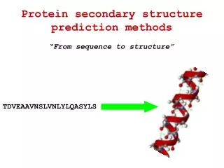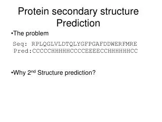
Secondary structure prediction
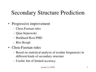
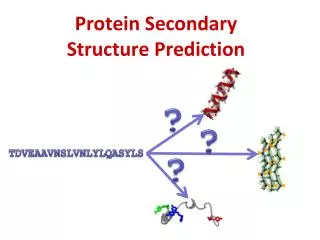
Secondary structure prediction. There have been many strategies since the 1970’s to predict secondary structure: – stereochemical principles (periodicity of helix, strands) – statistics (amino acid preference for helix/strand/coil) PSIPRED is one of the latest prediction methods

Secondary structure prediction
E N D
Presentation Transcript
Secondary structure prediction There have been many strategies since the 1970’s to predict secondary structure: – stereochemical principles (periodicity of helix, strands) – statistics (amino acid preference for helix/strand/coil) PSIPRED is one of the latest prediction methods – uses multiple alignments of sequences and structural information – not as computer intensive as its predecessors that also used databases The accuracy is > 70% against a known structure test dataset. This may be the practical limit of secondary structure prediction owing to the natural variation of secondary structures in homologous proteins that are used to train the prediction method.
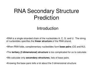
This PSIPRED prediction of a DNA binding domain is very close to reality. There are four helices in this domain. Only the boundaries vary from the actual structure
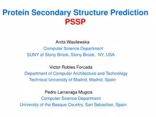
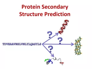
Secondary structure is useful to find folds A real life example ! I was interested in a protein called Coilin which serves as a scaffold or glue that helps proteins involved in RNA maturation to come together (splicing factors, telomerase, etc). I knew the C-terminal portion of Coilin had a folded domain in it by NMR spectroscopy. mystery Domain
Secondary structure is useful to find folds From PSIPRED, the mystery domain was predicted to have four strands and one small 3-10 helix as well as two large undefined regions Mystery domain
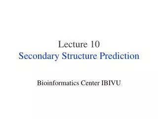
Secondary structure is useful to find folds I determined the structure of the Coilinmystery domain by NMR methods and observed a similar fold with two very large disordered loops Because the loops took up half of the protein domain’s sequence, primary sequence search methods such as BLAST could not find a homolog in the PDB (too many gaps, probably). However, a secondary search using SSEA did find many homologous folds that aided my structure determination. For example, I could build a 3D molecular model and then compare my NMR experimental observations to it.
Secondary structure is useful to find folds Some hits discovered by SSEA that had the same number of strands in the same order as the Coilin mystery domain I made molecular models of Coilin from these structures and then challenged my NMR experimental observations against them
Secondary structure is useful to find folds Tudor domains are known to binding methylated lysines and arginines in histones. Other related domains bind peptides and even DNA/RNA. They are very versatile. I performed biochemical experiments and ruled out every binding mode except for protein-protein interactions for the Coilin mystery domain.
Finding folds that are similar in the PDB This is a comparison of 3D coordinates versus all of the entries in the Protein Data Bank A program called SSM is very popular and fast - pairwise comparison and 3D alignment of protein structures - multiple comparison and 3D alignment of protein structures - examination of a protein structure for similarity with the whole archive
Finding folds that are similar in the PDB This protein was used as an example in the previous unit when I presented TOPS diagrams. It’s a viral protein from bacteriophage lambda that serves as the “collar” between the “head” and the “tail” of the virus gpU
Finding folds that are similar in the PDB Like the Coilin mystery domain, PSIPRED correctly predicted all of the beta strands and alpha helices in the protein. Here is the TOPS diagram of that prediction:
Finding folds that are similar in the PDB Despite having a very similar fold, a BLAST primary sequence search failed to detect any homologs in the Protein Data Bank, presumably to the extremely evolutionary distance between viruses and other representatives in the database. Once I performed a 3D SSM search for similar folds, I found these three, among others. My solved structure of gpU has the PDB accession number 1ZIZ.