Download
1 / 66
670 likes | 830 Views
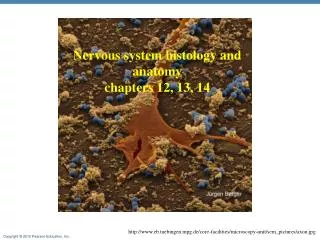
Nervous system histology and anatomy chapters 12, 13, 14. http://www.eb.tuebingen.mpg.de/core-facilities/microscopy-unit/sem_pictures/axon.jpg. Functions of the Nervous System. Sensory input Information gathered by sensory receptors about internal and external changes Integration

E N D
Nervous system histology and anatomy chapters 12, 13, 14 http://www.eb.tuebingen.mpg.de/core-facilities/microscopy-unit/sem_pictures/axon.jpg
Functions of the Nervous System • Sensory input • Information gathered by sensory receptors about internal and external changes • Integration • Interpretation of sensory input (CNS) • Motor output • Activation of effector organs (muscles and glands) produces a response
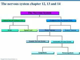
Anatomical Organization of the Nervous System Central nervous system (CNS) Brain and spinal cord Integration and command center Peripheral nervous system (PNS) Paired spinal and cranial nerves Carries messages to and from the spinal cord and brain
The major components and functions of the nervous system Central Nervous System The central nervous system (CNS) consists of the brain and spinal cord and is responsible for integrating, processing, and coordinating sensory data and motor commands. Information processing includes the integration and distribution of information in the CNS. Peripheral Nervous System The motor division of the PNS carries motor commands from the CNS to peripheral tissues and systems. The peripheral nervous system (PNS) includes all the neural tissue outside the CNS. includes The somatic nervous system (SNS) controls skeletal muscle contractions. The autonomic nervous system (ANS) provides automatic regulation of smooth muscle, cardiac muscle, glands, and adipose tissue. The sensory division of the PNS brings information to the CNS from receptors in peripheral tissues and organs. Special sensory receptors provide sensations of smell, taste, vision, balance, and hearing. Somatic sensory receptors provide position, touch, pressure, pain, and temperature sensations. • Smooth muscle • Cardiac muscle • Glands • Adipose tissue Skeletal muscle Visceral sensory receptors monitor internal organs. Effectors are target organs whose activities change in response to neural commands. Receptors are sensory structures that detect changes in the internal or external environment. Figure 11 Section 1
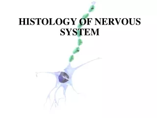
Histology of Nerve Tissue The two cell types of the nervous system are: Neurons– excitable cells that transmit electrical signals Supporting cells – cells that surround and wrap neurons
Neurons (Nerve Cells) Structural units of the nervous system Composed of a body, axon, and dendrites Long-lived (over 100 years) Amitotic – once achieve there roles in the system they loose the ability to divide High metabolic rate – require high supply of oxygen and glucose Their plasma membrane function in Electrical signaling
Neuron Cell Body (Perikaryon or Soma) Posses all the components needed to produce membrane parts, synthesize enzymes and neurotransmitters: Contains the nucleus and a nucleolus rough endoplasmic reticulum/Nissle bodies for protein synthesis Golgi apparatus which packages materials into vesicles for transport Numerous mitochondria Cytoskeletal elements Has no centrioles (hence its amitotic nature) Neural stem cells are still found in the adult but are not active Cells in the hippocampus (part that is involves memory) can still dividing
Dendrites Short branched processes They are the receptive, or input, regions of the neuron Supply big surface for receiving signals Convey incoming messages towards the cell body. Electrical signals are conveyed as graded potentials (not action potentials)
Axons: Structure The axon contains the same organelles as the cell body with the exception of Nissle bodies and Golgi apparatus (protein synthesis and packaging) Depends on the cell body to supply proteins Each neuron has single axon of uniform diameter Initiate in an enlarged area called the axon hillock Numerous terminal branches (telodendria) Knoblike axon terminals (synaptic knobs or boutons) Secretory region of neuron Release neurotransmitters to excite or inhibit other cells Long axons are called nerve fibers
Axons: Function Generate and transmit action potentials – away from the cell The signal starts at the junction between the axon hillock and axon – trigger zone Secrete neurotransmitters from the axonal terminals in response to the impulse arriving along the axon Movement along axons (not the signal movement) occurs in two ways Anterograde — toward axonal terminal (mitochondria, cytoskeletal elements, membrane components for membrane renewal, enzymes Examples: mitochondria, membrane components, enzymes Retrograde — away from axonal terminal (organelles to be degraded, signal molecules, viruses, and bacterial toxins like polio, rabies, herpes simplex)
Supporting Cells: Neuroglia Two groups In the CNS – astrocytes, microglia, ependymal cells and oligodendrocytes In the PNS – satellite and Schwann cells
Neuroglia in the CNS: Astrocytes Most abundant, versatile, and highly branched glial cells In some areas of the brain they account for 90% of the tissue They cling to neurons and their synaptic endings, and cover capillaries Capillary Neuron Astrocyte http://www.ucl.ac.uk/biology/images/astrocytes.gif Figure 11.3a
Neuroglia in the CNS: Astrocytes functions Maintaining blood-brain barrier – preventing free access of circulating compounds to the CNS. The extensions of the astrocytes end in expanded portion that wraps capillaries
Why are the capillaries in the BBB less permeable? • Endothelial cells form tight junctions that prevent solutes movement between cells • Astrocytes • Selective transport properties of the endothelial cells • The BBB • Helps maintain a stable environment for the brain • Separates neurons from some bloodbornesubstances
Blood-Brain Barrier: Functions • Selective barrier that allows nutrients to pass freely • Is ineffective against substances that can diffuse through plasma membranes (ex. Ethanol, caffeine) • Absent in some areas: • Ex. - hormones generally do not penetrate the brain from the blood, so in order to control the rate of hormone secretion effectively, there are specialized sites where neurons can "sample" the composition of the circulating blood. At these sites, the blood-brain barrier is 'leaky‘ (pituitary gland) • Capillaries of the choroid plexus • The BBB can break down under certain conditions: • hypertension, radiation, infection and brain trauma
Example • In Parkinson’s disease there is a lack in the NT dopamine (neurons that produce it are either damaged or dead) • Dopamine can not be given in a pill or injection because it can’t cross the BBB • Instead, a precursor - L-dopa – is given. This is being transported across the BBB and is being used by neurons
Neuroglia in the CNS: Astrocytes functions • Creating a framework for the CNS with microfilaments that extend from the astrocytes • Repairing damaged neural tissue- limited structural repairs that stabilize and prevent further injury. • In the embryonic brain, the astrocyte appear to be involve in directing the growth and interconnection of developing neurons. • Secrete nerve growth factors that promote neuron growth and synapse formation
Neuroglia in the CNS: Astrocytes functions Control the chemical environment Regulating the concentration of sodium, potassium and carbon dioxide ions Providing system for transporting nutrients and dissolved gasses between capillaries and neurons absorb neurotransmitters to prevent excessive levels in tissue fluid; control ion concentration in the interstitial fluid
Myelin sheath Process of oligodendrocyte Nerve fibers Oligodendrocytes • Branched cells • Processes wrap CNS nerve fibers, forming insulating myelin sheaths
Neuroglia cells in the PNS Schwann cells (neurolemmocytes) – surround fibers of the PNS Similar function as the oligodendrocytes Satellite cells surround neuron cell bodies in the PNS similar function to the astrocytes in the CNS – controlling the chemical environment
Satellite cells Cell body of neuron Schwann cells (forming myelin sheath) Nerve fiber (e) Satellite cells and Schwann cells (whichform myelin) surround neurons in the PNS. Figure 11.3e
Myelin Sheath: Formation Formed by Schwann cells in the PNS Schwann cells wraps many times around the axon Myelin sheath — concentric layers of Schwann cell membrane Neurilemma—peripheral bulge of Schwann cell cytoplasm Myelin sheath functions : Protect the axon Electrically insulate fibers from one another Increase the speed of nerve impulse transmission
Nodes of Ranvier (Neurofibral Nodes) Gaps in the myelin sheath between adjacent Schwann cells They are the sites where axon collaterals can emerge http://www.jdaross.cwc.net/introd13.jpg
Unmyelinated Axons • Thin nerve fibers are unmyelinated • One Schwann cell may incompletely enclose 15 or more unmyelinated axons
Unmyelinated Axons A Schwann cell surrounds nerve fibers but coiling does not take place Schwann cells partially enclose 15 or more axons http://www.bu.edu/histology/p/21201loa.htm
Axons of the CNS Both myelinated and unmyelinated fibers are present Myelin sheaths are formed by oligodendrocytes Nodes of Ranvier are widely spaced
Regions of the Brain and Spinal Cord White matter – dense collections of myelinated fibers Gray matter – mostly soma (cell bodies) and unmyelinated fibers
CNS: Gray and White Matter Figure 9.5a
Neurons Cell body locations Groups of cell bodies are named according to their location: Most cell bodies are in the CNS in clusters called nuclei Some are in the PNS in clusters called ganglia
Neurons’ Processes location The groups of axons are named according to their locations: In the CNS the extensions called tracts In the PNS the extensions (axons) are called nerves
Neuron Classification - Structural Multipolar — three or more processes Many extensions; many dendrites lead toward cell body, one axon leads away from cell body. Most neurons in the CNS and those whose axons carry impulses away from the CNS are multipolar. Bipolar — two processes (axon and dendrite) Two extensions; one fused dendrite leads toward cell body, one axon leads away from cell body These are rare and can be found as part of the receptor apparatus in the eye, ear and olfactory mucosa. Unipolar — single, short process One very short process from cell body that divides into central and peripheral processes. Only the distal ends of the peripheral process are dendrites and the rest act as an axon along with the central process. Nearly all neurons that conduct impulses towards the CNS are unipolar.
Neuron Classification - Functional Sensory (afferent) - transmit impulses towardthe CNS Most are unipolar neurons with their bodies in ganglia in the PNS. Location – sensory receptors in the internal organs, skin, skeletal muscles and joints. They sense changes in the immediate environment Motor (efferent) - carry impulses away from the CNS Carry activating impulses from CNS and to the viscera, body muscles and glands; They are multipolar and their cell bodies are in the CNS.
Neuron Classification - Functional Interneurons (association neurons) - shuttle signals through CNS pathways Link other neurons together (i.e. sensory neuron to interneuron to motor neuron). All bodies are in CNS and they are multipolar.
Central nervous system http://www.courses.vcu.edu/DANC291-003/unit%201.htm
3 Major levels of the CNS • Spinal cord • involved in walking movement, reflexes of both skeletal muscles and the autonomic nervous system effectors • Lower brain/subcortical level • Subconscious activities of both the autonomic and somatic nervous system. Involved in several emotional patterns • Higher brain/cortical level • Very large memory storage, place of higher functions like thoughts, awareness
CNS protection • Bony protection by the skeleton – brain by skull and spinal cord by vertebrae • 3 layers of membrane collectively called meninges that are found between the bone and the CNS tissue • Cerebro-spinal fluid – CSF - found in the ventricles, central canal and subarachnoid space • Blood brain barrier - BBB
CNS: Physical Support Figure 9.2a
CNS: Physical Support Figure 9.2b
lateral 3 4 Ventricles • Filled with CSF (cerebrospinal fluid) • Ventricles continuous w/each other + central canal of spinal cord • Lateral Ventricle (#1+2) • Cerebral Hemisphere • Third Ventricle • Diencephalon • Fourth Ventricle • Cerebral Aqueduct: connects 3rd and 4th ventricles • Connects to central canal of spinal cord & medulla
Choroid Plexuses and CSF • Clusters of capillaries and the ependymal cells http://www.sci.uidaho.edu/med532/choroid.htm
CSF production • The choroid plexus forms tissue fluid filters • Have ion pumps that selectively pump ions from the plasma into the ventricles. • The ions pumps create osmotic pressure that draws water to the CSF • the choroid plexus of the lateral ventricles producing the most. • The rate of formation is approximately 0.35 ml/min or 500 ml/day; a rate which replaces the total volume of CSF approximately 2-3 times over in 24 hours.
CSF – cerebrospinal fluid functions • Liquid cushion for brain and spinal cord • Nourishes brain • Removes waste • Conducts chemical signals between parts of CNS
Brain: Midsagittal View Cerebrum Corpus callosum Forebrain Thalamus Diencephalon Hypothalamus Pituitary gland Cerebellum Midbrain Brainstem Pons Spinal cord Medulla oblongata (c) Midsagittal section Figure 9.11c
Basal nuclei Cerebrum functional regions • The cerebrum has three basic regions: cortex (gray matter), white matter, and basal nuclei (gray matter) http://www.bioon.com/book/biology/whole/image/1/1-6.tif.jpg
Layers of the Cerebral Cortex Figure 9.12a–b
Functional Areas of Cerebrum Primary motor cortex (voluntary movement) Central sulcus Premotor cortex (coordinates voluntary movements) Primary somatosensory cortex (somesthetic sensations and proprioception) Sensory association areas (integration of sensory information) Prefrontal association areas (idea and plan for voluntary movement, thoughts, personality) Visual association areas (higher vision processing) Primary visual cortex (vision) Broca’s area (speech formation) Wernicke’s area (language comprehension) Olfactory cortex (smell) Limbic association cortex (emotions, learning, and memory) Auditory association areas Primary auditory cortex (hearing) Figure 9.14
Beneath the cerebrum – basal nuclei • Are masses of gray matter that are embedded in white matter of cerebrum • Function in the subconscious control of skeletal muscle tone • The coordination of learned movement patterns (walking, lifting)
White Matter of the Cerebrum • Association fibers • Connections within one hemisphere • Commissural fibers connecting two hemispheres: • corpus callosum • anterior commissure • Projection fibers link cerebral cortex with: • diencephalon, brain stem, cerebellum, and spinal cord
Diencephalon • Consists of three structures • thalamus • Hypothalamus • epithalamus • Encloses the third ventricle