
Osteological Overview of Pelvis and Femur
E N D
Presentation Transcript
HIP Dr. Michael P. Gillespie
OSTEOLOGY • Each innominate is the union of three bones: the ilium, pubis, and ischium. • The right and left innominates connect with each other anteriorly at the pubic symphysis and posteriorly at the sacrum. • An osteoligamentous ring known as the pelvis (Latin: basin or bowel) is formed. • Functions of the pelvis: • Attachment point for many muscles of the lower extremity and trunk. • Transmits the weight of the upper body and trunk to the ischial tuberosities during sitting and to the lower extremities during standing or walking. • Supports the organs of the bowel, bladder, and reproductive system. Dr. Michael P. Gillespie
INNOMINATE • Ilium • Pubis • Ischium • Acetabulum Dr. Michael P. Gillespie
EXTERNAL SURFACE OF THE PELVIS • Wing (ala) – the large fan-shaped wing of the ilium forms the superior half of the innominate. • Acetabulum – a deep, cup-shaped cavity below the wing. • Obturator-foramen – the largest foramen in the body. Covered by the obturator membrane. Dr. Michael P. Gillespie
OSTEOLOGIC FEATURES OF THE ILIUM • External Surface • Posterior, anterior, and inferior gluteal lines • Anterior-superior iliac spine • Anterior-inferior iliac spine • Iliac crest • Posterior-superior iliac spine • Posterior-inferior iliac spine • Greater sciatic notch • Greater sciatic foramen • Sacrotuberous and sacrospinous ligaments • Internal Surface • Iliac fossa • Auricular surface • Iliac tuberosity Dr. Michael P. Gillespie
OSTEOLOGIC FEATURES OF THE PUBIS • Superior pubic ramus • Body • Crest • Pectineal line • Pubic tubercle • Pubic symphysis joint and disc • Inferior pubic ramus Dr. Michael P. Gillespie
OSTEOLOGIC FEATURES OF THE ISCHIUM • Ischial spine • Lesser sciatic notch • Lesser sciatic foramen • Ischial tuberosity • Ischial ramus Dr. Michael P. Gillespie
ANTERIOR ASPECT: PELVIS, SACRUM, RIGHT PROXIMAL FEMUR Dr. Michael P. Gillespie
LATERAL VIEW RIGHT INNOMINATE BONE Dr. Michael P. Gillespie
POSTERIOR ASPECT OF PELVIS, SACRUM, & PROXIMAL FEMUR Dr. Michael P. Gillespie
FEMUR • The longest and strongest bone in the human body. • The femoral head projects medially and slightly anterior to articulate with the acetabulum. • The femoral neck connects the head with the shaft. • The neck displaces the proximal shaft of the femur laterally away from the joint, thereby reducing the likelihood of bony impingement. • Distal to the neck, the shaft of the femur courses slightly medially, placing the knees and feet closer to the midline of the body. • The femur bows slightly when subjected to the weight of the body. Stress along the bone is dissipated through compression along the posterior shaft and through tension along the anterior shaft. Dr. Michael P. Gillespie
OSTEOLOGIC FEATURES OF THE FEMUR • Femoral Head • Femoral Neck • Intertrochanteric Line • Greater trochanter • Trochanteric fossa • Intertrochanteric crest • Quadrate tubercle • Lesser trochanter • Linea aspera • Pectineal (spiral) line • Gluteal tuberosity • Lateral and medial supracondylar lines • Adductor tubercle Dr. Michael P. Gillespie
ANTERIOR ASPECT RIGHT FEMUR Dr. Michael P. Gillespie
MEDIAL & POSTERIOR SURFACES RIGHT FEMUR Dr. Michael P. Gillespie
ANGLE OF INCLINATION • The angle of inclination of the proximal femur describes the angle within the frontal plane between the femoral neck and the medial side of the femoral shaft. • At birth this angle is about 140 – 150 degrees; however, the loading across the femoral neck during walking usually decreases this to the normal adult value of about 125 degrees. • Coxa = hip, vara = to bend inward, valga = to bend outward • Coxa vara – an angle of inclination markedly less than 125 degrees. • Coxa valga – an angle of inclination markedly greater than 125 degrees. • Abnormal angles can lead to dislocation or stress-induced degeneration of the joint. Dr. Michael P. Gillespie
ANGLE OF INCLINATION Dr. Michael P. Gillespie
FEMORAL TORSION • Femoral torsion describes the relative rotation (twist) between the bone’s shaft and neck. • Normally, as viewed from above, the femoral neck projects about 15 degrees anterior to a mid-lateral axis through the femoral condyles (normal anteversion). • Femoral torsion significantly different than 15 degrees is considered abnormal. • Excessive anteversion – significantly greater than 15 degrees • Retroversion – approaching 0 degrees • Healthy infants are born with about 40 degrees of femoral anteversion Dr. Michael P. Gillespie
EXCESSIVE FEMORAL ANTEVERSION • Excessive anterversion that persists into adulthood can increase the likelihood of hip dislocation, articular incongruence, increase joint contact force, and increased wear on the cartilage. • This can lead to secondary osteoarthritis of the hip. • It may be associated with an abnormal gait pattern called “in-toeing”, a walking pattern with exaggerated posturing of hip internal rotation. • The amount of “in-toeing” is generally related to the amount of femoral anteversion. • It is a compensatory mechanism used to guide the excessively anteverted femoral head more directly into the acetabulum. • Over time, shortening of the internal rotator muscles and ligaments occurs, thereby reducing external rotation. • Most children with in-toeing eventually walk normally. • Excessive femoral anteversion is common in persons with cerebral palsy. It typically does not resolve in this population. Dr. Michael P. Gillespie
NORMAL ANTEVERSION Dr. Michael P. Gillespie
EXCESSIVE ANTEVERSION Dr. Michael P. Gillespie
RETROVERSION Dr. Michael P. Gillespie
INTERNAL ROTATION IMPROVING JOINT CONGRUITY Dr. Michael P. Gillespie
IN-TOEING Dr. Michael P. Gillespie
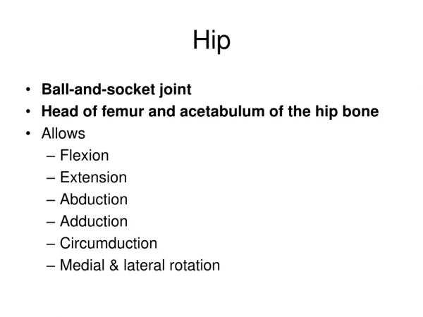
FUNCTIONAL ANATOMY OF THE HIP JOINT • The hip is a classic ball-and-socket joint secured within the acetabulum by an extensive set of connective tissues and muscles. • Articular cartilage, muscle, and cancellous bone in the proximal femur help dampen the large forces that cross the hip. Dr. Michael P. Gillespie
FEMORAL HEAD • The head of the femur forms about two-thirds of a nearly perfect sphere. • The entire surface of the femoral head is covered by articular cartilage except for the region of the fovea, which is slightly posterior to the center of the head. • The fovea is a prominent pit that serves as the attachment point for the ligamentum teres. • The ligamentum teres is a tubular sheath that runs between the transverse acetabular ligament and the fovea of the femoral head. It is a sheath that contains the acetabular artery. Dr. Michael P. Gillespie
ACETABULUM • The acetabulum (Latin – vinegar cup) is a deep, hemispheric cuplike socket that accepts the femoral head. • The femoral head contacts the acetabulum along the horseshoe-shaped lunate surface, which is covered with thick articular cartilage. • During walking, hip forces fluctuate from 13% of body weight to over 300% of body weight during the mid-stance phase. • During stance phase, the lunate surface flattens slightly as the acetabular notch widens. This serves as a dampening mechanism to reduce peak pressure. • The acetabular fossa is a depression located deep within the floor of the acetabulum. It does not normally come into contact with the femoral head. Dr. Michael P. Gillespie
HIP JOINT COMPRESSION AS A PERCENT OF GAIT CYCLE Dr. Michael P. Gillespie
ANATOMIC FEATURES OF THE HIP JOINT • Femoral Head • Fovea • Ligamentum teres • Acetabulum • Acetabular notch • Lunate surface • Acetabular fossa • Labrum • Transverse acetabular ligament Dr. Michael P. Gillespie
INTERNAL ANATOMY OF HIP JOINT Dr. Michael P. Gillespie
ACETABULAR LABRUM • The acetabular labrum is a flexible ring of fibrocartilage that surrounds the outer circumference (rim) of the acetabulum. • The acetabular labrum projects about 5 mm toward the femoral head. • It provides significant stability to the hip by “gripping” the femoral head and deepening the volume of the socket by approximately 30%. • The seal formed by the labrum maintains a negative intra-articular pressure, thereby creating a modest suction that resists distraction of the joint surfaces. • It also helps to hold synovial fluid within the joint space. • It decreases the contact stress (force / area) by increasing the surface area of the acetabulum. • Poor blood supply – limited ability to heal • Well supplied with afferent nerves – proprioceptive feedback / pain Dr. Michael P. Gillespie
ACETABULAR ALIGNMENT • In the anatomic position, the acetabulum typically projects laterally from the pelvis with a varying amount of inferior and anterior tilt. • Congenital or developmental conditions can result in an abnormally shaped acetabulum. • A dysplastic acetabulum that does not adequately cover the femoral head can lead to chronic dislocation and increased stress, which can lead to osteoarthritis. • Two measurements are used to describe the extent to which the acetabulum naturally covers and helps to secure the femoral head: • Center-edge angle • Acetabular anteversion angle Dr. Michael P. Gillespie
CENTER-EDGE ANGLE • The center-edge angle varies widely, but on average measures about 35 degrees in adults. • A significantly lower center-edge angle reduces the acetabular coverage of the femoral head. This increases the risk of dislocation and reduces contact area within the joint. • During the single-limb-support phase of walking, this reduced surface area would increase joint pressure (force / area) by about 50%. • This increased joint pressure can lead to premature osteoarthritis. Dr. Michael P. Gillespie
CENTER-EDGE ANGLE Dr. Michael P. Gillespie
ACETABULAR ANTEVERSION ANGLE • The acetabular anteversion angle measures the extent to which the acetabulum projects anteriorly within the horizontal plane, relative to the pelvis. • Observed from above, the normal acetabular anteversion angle is about 20 degrees, which exposes part of the anterior side of the femoral head. • A hip with excessive acetabular anteversion is more exposed anteriorly. • When anteversion is severe, the hip is more prone to anterior dislocation and associated lesions of the labrum. Dr. Michael P. Gillespie
ACETABULAR ANTEVERSION ANGLE Dr. Michael P. Gillespie
CAPSULE AND LIGAMENTS OF THE HIP • A synovial membrane lines the internal surface of the hip joint capsule. • The iliofemoral, pubofemoral, and ischiofemoral ligaments reinforce the external surface of the capsule. • Passive tension in the stretched ligaments, the adjacent capsule, and the surrounding muscles help to define end-range movements of the hip. • Increasing the flexibility of parts of the capsule is an important component of manual physical therapy for restricted movement of the hip. Dr. Michael P. Gillespie
ANTERIOR CAPSULE & LIGAMENTS Dr. Michael P. Gillespie
POSTERIOR CAPSULE & LIGAMENTS Dr. Michael P. Gillespie
PARAPLEGIC WITH SUPPORT BRACES Dr. Michael P. Gillespie
TISSUES THAT BECOME TAUT AT THE END-RANGES OF PASSIVE HIP MOTION Dr. Michael P. Gillespie
TISSUES THAT BECOME TAUT AT THE END-RANGES OF PASSIVE HIP MOTION Dr. Michael P. Gillespie
CLOSE-PACKED POSITION OF THE HIP • Full extension of the hip (about 20 degrees beyond neutral) in conjunction with slight internal rotation and slight abduction twists or “spirals” the fibers of the capsular ligaments to their most taut position. • This is considered the close-packed position of the hip. • The passive tension leads to stability of the joint and reduces “joint play”. • The hip joint is one of the few joints in the body where the close-packed position is NOT also the position of maximal joint congruency. They fit most congruently in about 90 degrees of flexion, moderate abduction, and external rotation. Dr. Michael P. Gillespie
NEUTRAL AND CLOSED PACKED POSITIONS Dr. Michael P. Gillespie
OSTEOKINEMATICS • Reduced hip motion may be an early indicator of disease or trauma. • Limited hip motion can impose functional limitations on activities such as walking, standing upright, or picking up objects on the floor. • Femoral-on-pelvic hip osteokinematics – rotation of the femur about a relatively fixed pelvis. • Pelvic-on-femoral hip osteokinematics – rotation of the pelvis, and often the superimposed trunk, over relatively fixed femurs. • Movements: • flexion & extension in the sagittal plane, abduction & adduction in the frontal plane, and internal and external rotation in the horizontal plane. • The anatomic position is the 0-degree or neutral reference point. Dr. Michael P. Gillespie
FEMORAL-ON-PELVIC OSTEOKINEMATICS • Rotation of the Femur in the Sagittal Plane • Hip flexion to 120 degrees • Full knee extension limits hip flexion to 70 – 80 degrees due to increased tension in the hamstrings • Hip extension to 20 degrees • Full knee flexion reduces hip extension due to tension in rectus femoris • Rotation of the Femur in the Frontal Plane • Hip abduction to 40 degrees • Limited by pubofemoral ligament and adductors • Hip adduction to 25 degrees • Limited by interference with contralateral limb, passive tension in hip abductors, iliotibial band, and ischiofemoral ligament • Rotation of the Femur in the Horizontal Plane • Internal rotation to 35 degrees • Produces tension in piriformis and ischiofemoral ligament • External rotation to 45 degrees • Produces tension in internal rotators and iliofemoral ligament Dr. Michael P. Gillespie
SAGITTAL PLANE ROTATIONS Dr. Michael P. Gillespie
FRONTAL PLANE ROTATIONS Dr. Michael P. Gillespie
HORIZONTAL PLANE ROTATIONS Dr. Michael P. Gillespie
FEMORAL-ON-PELVIC (HIP) MOTION Dr. Michael P. Gillespie
PELVIC-ON-FEMORAL OSTEOKINEMATICS • Pelvic rotation in the Sagittal Plane • Pelvic rotation in the Frontal Plane • Pelvic Rotation in the Horizontal Plane Dr. Michael P. Gillespie