Download
1 / 35
360 likes | 395 Views
Explore the functions of the urinary system in maintaining homeostasis, with a focus on nephrons, glomerular filtration, and the renal anatomy.

E N D
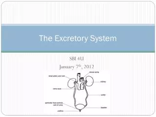
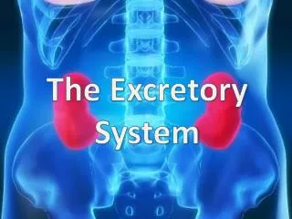
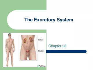
The Excretory System Chapter 23
What is the Urinary System? • The purifying system for blood. • Blood passes through the urinary system in order to be cleaned of toxins and poisons. • Organs of the urinary system include: Kidneys – paired Ureters – paired Bladder – single Urethra - single
The human excretory system regulates the chemical composition of body fluids by removing metabolic wastes and retaining the proper amounts of water, salts, and nutrients. Components of this system in vertebrates include the kidneys, liver, lungs, and skin.
By doing this, the excretory system allows the body to maintain HOMEOSTASIS. • Homeostasis is the body’s ability to maintain a constant internal environment.
The Urinary System • The KIDNEYS are found approximately 2 inches from the vertebral column (3 fingerbreadths lateral). • The right kidney is LOWER than the left. • The superior edge of the left kidney is at T12. The inferior edge of the right is at L3. • They are moveable during breathing. They move approximately I inch in either direction.
The URETER enters the bladder from below. Which means that the bladder fills from the bottom. • The URETHRA is central. It is approx. 1.5 inches long in females and 8 inches long in males. This fact is important because the length of a foley catheter differs in the sexes.
A coronal section (frontal section) of the kidney will reveal the junction of the ureter and kidney form a funnel. • This area is called the RENAL PELVIS. The renal pelvis contains the COLLECTING TUBULES from the RENAL MEDULLA.
The outer portion of the kidney is the RENAL CORTEX. In humans, the renal cortex extends between the RENAL PYRAMIDS. • Each pyramid branches into smaller funnels called CALYX. Each calyx receives part of the medulla. Each calyx has its own pyramid.
The Nephron The nephron consists of a cup-shaped capsule containing capillaries and the glomerulus, and a long renal tube. Blood flows into the kidney through the renal artery, which branches into capillaries associated with the glomerulus. Arterial pressure causes water and solutes from the blood to filter into the capsule. Fluid flows through the proximal tubule, which include the loop of Henle, and then into the distal tubule. The distal tubule empties into a collecting duct. Fluids and solutes are returned to the capillaries that surround the nephron tubule.
The nephron has three functions: • Glomerular filtration of water and solutes from the blood. • Tubular reabsorption of water and conserved molecules back into the blood. • Tubular secretion of ions and other waste products from surrounding capillaries into the distal tubule.
How do the nephrons work? 1. The outermost layer of the kidneys, the cortex, is composed of approximately 1,250,000 structural units called nephrons. Blood is carried to the kidneys by the renal arteries, which branch into smaller arteries inside the cortex and then lead to clusters of capillaries called glomeruli.
2. Each glomerulus is surrounded by a "C"-shaped structure called the Bowman's Capsule. It is here that materials such as urea, salts, water, glucose, & others pass from the blood into the nephron.
3. These materials (referred to as the "filtrate") pass through the loop of Henle. As the filtrate travels through the loop of Henle, useful substances are reabsorbed into the surrounding capillaries (which connect to veins that will transport the "clean" blood back to the heart via the renal vein).
4. About 180 liters of filtrate is produced each day, but only 1.5 liters of urine. So as you can see, most materials that initially enter the nephron are reabsorbed, leaving only the urea, salts, & some water in the tubule. These metabolic wastes form urine, which is transported to the renal pelvis by the collecting tubule.
Renal capsule Minor calyx Major calyx Renal artery Renal vein Renal pelvis ureter Renal medulla Renal pyramid Renal cortex Renal column
Adult normal kidney with a renal cyst. A much larger cyst at the upper pole of the kidney
There are approximately 8-10 calyces and a pyramid for each one in each human kidney. • The functional unit of the kidney is the NEPHRON. There are about 2.5 million nephrons in each kidney. • The ADRENAL GLANDS (suprarenal glands) are endocrine glands found on the superior portion of each kidney. They secrete EPINEPHRINE and NOREPINEPHRINE.
The Layers of the Kidney • Each layer has a specific job 1. Cortex - nephrons for filtration 2. Medulla – collecting layer 3. Pelvis – collecting tubules come together
Suprarenal glands Suprarenal glands Kidney Renal vessels urachus urachus Is this a male or female? Trigone ureter Bladder Trigone of bladder Prostatic Urethra Prostate Gland
Ureter 1 – epithelium 2 – connective tissue 3 – smooth muscle Lumen
What is in Urine? Urea – derived from the breakdown of amino acids (building blocks of proteins). Uric Acid – results from the turnover of nucleic acids. Creatinine – formed by the breakdown of creatine phosphate (found in muscle)
Other functions of the urinary system • Regulation of the volume and chemical makeup of the blood. • Maintenance of the proper balance between water and salts and between acids and bases.
Ureters • Carry urine from the kidneys to the bladder. • Begin as a continuation of the RENAL PELVIS in the kidney.
Bladder • A collapsible muscular sac that store urine. • Lies on the pelvic floor, posterior to the pubic symphysis. • The bladder lies anterior to the rectum in males. • In females, it lies anterior to the vagina and uterus. • Ureters enter the bladder at the POSTEROLATERAL ANGLES. • The apex of the bladder (anterior angle) has a fibrous band called the URACHUS (remnant of the embryonic urinary canal). • The inferior angle drains into the URETHRA. • In males, the PROSTATE GLAND lies directly inferior to the bladder surrounding the urethra.
Inside the bladder are the two openings where the ureters enter. When combined with the opening of the urethra, a triangle is formed known as the TRIGONE.
The wall of the bladder has three layers: • Mucosa – distensible epithelium • Thick Muscle (Detrusor Muscle) – intermingled smooth muscle fibers • Fibrous Adventitia
When empty, the bladder collapses into a pyramid shape. • As urine accumulates, the walls distend and stretch. There is no increase in internal pressure until there is about 300mL of accumulated urine. • A full bladder holds about 500mL of urine.
At the junction between the bladder and urethra, the DETRUSOR MUSCLE forms the INTERAL URETHRAL SPHINCTER. • The sphincter keeps the urethra closed when urine is not being passed. It prevents “dribbling”. Involuntary action – smooth muscle. • The EXTERNAL URETHRAL SPHINCTER surrounds the urethra in a sheet of muscle called the UROGENITAL DIAPHRAGM. • The EUS is what humans use to voluntarily delay urination. Therefore, it is skeletal muscle.
The urethra is of different lengths in each sex. • Males – 20 cm long. Has three regions: a. Prostatic urethra b. Membranous urethra – in the urogenital diaphragm. c. Penile urethra (spongy) • The urethra opens into the EXTERNAL URETHRAL ORIFICE.