Download
1 / 73
780 likes | 1.53k Views
Congenital Anomalies By Dr Shewikar Farrag. General objective. To identify different congenital anomalies that are present at birth. Specific objectives:. List the possible causes of fetal malformations. Define some common congenital anomalies in the newborn infant.

E N D
General objective • To identify different congenital anomalies that are present at birth.
Specific objectives: • List the possible causes of fetal malformations. • Define some common congenital anomalies in the newborn infant. • Describe some of the surgery related differences between infants and adults. • Outline important aspects in the pre and post operative care of paediatric patients. • Discuss specific pre and post operative care of a child with the following: cleft lip, cleft palate, oesophageal atresia and pyloric stenosis.
Causes of fetal malformation: • Drugs • Radiation • Viruses • Genetic traits
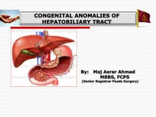
Common congenital anomalies in the newborn • Respiratory system: - Laryngeal stridor - Choanal atresia • Gastrointestinal system: - Anomalies of the mouth (cleft lip & cleft palate). - Anomalies of the esophagus (esophageal atresia & chalasia of the esophagus). • Anomalies of the stomach and duodenum: - pyloric stenosis - duodenal obstruction - hiatus hernia • Anomalies of the intestine: - Imperforated anus. - Omphalocele. - Intestinal atresia. - Diaphragmatic hernia. - Hirschsprung’s disease (congenital aganglionic megacolon) - Intussusception
Congenital anomalies of the urinary system: • Epispadias • Hypospadias • Phimosis • Hydrocele • Inguinal hernia • Polycystic kidney • Wilm’s Tumor (Embryoma)
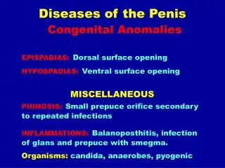
Epispadias • Mutual opening located on dorsal or superior surface of the penis.
Hypospadias • Urethral opening located behind glands penis or anywhere along ventral (lower) surface of penis shaft.
N.B • Infants with epispadias and hypospadias should not be circumcised before repair of the defect because the surgeon may wish to use a portion of the foreskin for plastic repair.
Phimosis • Narrowing or stenosis of preputial opening of foreskin. (in severe cases: circumcision or vertical division and transverse, suturing of foreskin)
Hydrocele • Fluid in scrotum. Therapeutic management is surgical repair indicated if spontaneous resolution not accomplished in 1 year.
Inguinal hernia: • Protrusion of abdominal contents through inguinal canal into the scrotum. Therapeutic management includes: detected as painless inguinal swelling of variable size surgical closure of inguinal defect.
Polycystic kidney: The infant has enlarged kidneys filled with cysts at birth If the condition is bilateral, the infant will not pass urine but if it is unilateral the condition may be missed until later in life.
Wilm’s Tumor (Embryoma) • It is a malignant tumor of the kidney that arises from an embryonic structure present in the child before birth; The tumor is felt as an abdominal mass. It is important that the necessary for diagnosis because handling appears to increase the danger of metastasis.
Skeletal defects affecting the nervous system: • Spina Bifida • Spina Bifida Occulta • Meningocele • Meningomyelocele • Hydrocephalus
Spina Bifida • It is a defective closure of the vertebral column. It is more common in the lumbo sacral region. It has varying degree of tissue protrusion through the bony cleft.
Spina Bifida Occulta Usually the 5th lumber and 1st sacral vertebrae are affected with no protrusion of interspinal contents the spinal cord and its cover the skin over the defect may reveal a dimple, small fatty mass or a tuft of hair.
Meningocele Is a protrusion through the spina bifida, which forms a soft, saclike appearance along the spinal axis and contains spinal fluid and meninges within the sac and covered with skin.
Meningomyelocele Is a more serious defect in which the spinal cord and / or nerve roots as well as meningocele covering protrude through the spina bifida. The degree and extent of neurogenic defect depend on the level of the defect. The higher the level the greater the defect. If in the lumbosacral, the usual of the defect is associated with a flaccid paralysis of the lower extremities, absent sensation to the level of the lesion and loss of bowel and bladder control.
Hydrocephalus The abnormal increase in cerebrospinal fluid volume within the intracranial cavity due to a defect in the cerebrospinal fluid drainage system, intracranial pressure increases, the scalp veins dilate, and the cranial suture begin to separate.
Orthopedic Anomalies: • Clubfoot: flexion at the ankle with inversion of the heel and fore foot. • Torticollis: is a condition in which there is a lateral inclination and a rotation of the head away from the midline of the body with limitation of the range of motion of the neck. • Congenital dislocation of the hip: in this condition the femur head is completely dislocated from the acetabulum. The infant shows limited ability to abduct the hip, asymmetry of the gluteal skin folds and inguinal creases, and shortening of the affected leg.
Surgery related differences between young children and adults: • The metabolic rate of the infant and young children is much greater proportionately than that of adult. Children are growing and need to be fed more frequently. • The body tissues of the child heal quickly because of his rapid rate of metabolism and growth. • The child usually needs proportionately less analgesic than adult patient to obtain relative comfort after surgical procedures. • The child lacks the reserve physical resources that are available to the adult. His general condition may change very rapidly. • Abnormal fluid loss is more serious in the infant and young child than in the adult. Fluid intake and out-put must be calculated very carefully.
General aspects of pre and post operative paediatric care: Critically ill newborn babies need to be transported to medical centres or paediatric hospitals. Transfer of those babies need to be safe to avoid any deterioration of the infant’s condition.
Transportation of the newborn: • Portable incubator with available oxygen supply. • Equipment for suctioning. • Paediatric nurse should be available during the transfer. • All pertinent infant information should accompany the infant as he goes from one health agency to another.
Pre-operative care: • Psychological preparation of the child. • Except in emergency situations, children should preferably be free from respiratory complications and signs of malnutrition. • Nothing per mouth should be provided to the child pre-operatively (duration depends on child’s age). • The incision over or the part involved in surgery must be washed and inspected. Shaving may be needed. • The mouth should be checked for loose teeth or for dentures (particularly in children of 6-8 yrs old). Any missing teeth should be charted in child’s record. • Remove batteries and pins from the child’s hair.
Clothing should be warm and loose. The child should be dressed in a hospital gown and under pants only. • Check the child’s identification band to see that is eligible and secure. • Pre-medication: sedatives and analgesics are usually given 2 hrs before surgery except in emergencies. • Urination and bowel movements should be charted (enemas are not done routinely unless required). • Nostrils should be cleaned before surgery. • Allow the child to keep his toy till he is under the aesthetic. • Parents should be allowed to accompany their children to the operation site if they so desire.
Post-operative care: • Vital signs • Airway patency • Warm cot or incubator • Side-lying position • Condition and placement of dressing • Check and mark any apparent drainage from wound • IV fluids monitoring (rate, possible infiltration) • Proper handling of the child • Right use of restraints • Urinary catheter care • Skin colour and temprature • Signs of shock • Time of starting oral fluids • Diet modification according to child’s age • Use of sedatives as prescribed • Encouragement of early ambulation when appropriate
Anomalies of the mouth • Cleft lip • Cleft palate
Definition of cleft lip: • A cleft lip is an abnormal opening in the middle of the upper lip. • A cleft lip is a separation of the two sides of the lip. • It usually looks like a gap in the skin of the upper lip. • It is a birth defect. • It is the most common birth defect of the head and face. • It can happen on one side of the lip (unilateral cleft lip) or both sides of the lip (bilateral cleft lip).
What causes it? • We do not know what causes cleft lip. • Studies show that it could be caused by both: • Genes • Environment during pregnancy • Drugs • Infections or illnesses • Smoking • Drinking
Definition of cleft palate: • A cleft palate is an opening in the roof of the mouth (palate).
Clinical manifestations: • Observable defects
Cleft lip repair is usually done within 6 to 12 weeks of age. • Cleft palate repair is generally postponed until later to take advantage of the palatal changes that occur with normal growth. • Most surgeons repair a cleft palate between 9 months to 1 year before the child develops faulty speech habits.
Diagnostic Evaluation: • Readily apparent by observation and palpation (cleft palate)
Objectives of therapeutic management: • Close defects surgically at the appropriate age. • Prevent the complications. • Habilitate for optimum use of residual impairments. • Facilitate normal growth and development of the child.
Nursing care plan for infant with cleft lip and /or palate repair:
Nursing Diagnosis: • Altered nutrition less than body requirements related to difficulty in eating.
Goal: • Nurse: provide adequate nutritional intake. • Patient: will receive optimum nutrition.
Interventions: • Administer diet appropriate for age (specify). • Modify feeding techniques to adjust to defect. • Hold the child in upright position. • Use special feeding appliances. • Bubble frequently. • Assist mother with breast-feeding if this is mother’s preference.
Expected outcome: • Infant consumes an adequate amount of nutrients (specify the amount). • Infant exhibits appropriate weight gain.
Nursing Diagnosis: • High risk for altered parenting related to infant with a highly visible physical defect.
Goal: • Nurse: facilitate family’s acceptance of infant. • Patient (family): will demonstrate acceptance.
Interventions: • Allow expression of feelings. • Convey attitude of acceptance of infant and family. • Indicate by behaviour that child is a valuable human being. • Describe results of surgical correction of defect (use photographs of satisfactory results). • Arrange meeting with other parents who have experiences of similar situations and coped successfully.
Expected outcome: • Family discusses feelings and concerns regarding child’s defect, its repair and future prospects. • Family exhibits an attitude of acceptance of infant.
Nursing care plan for infant with cleft lip and /or palate repair: