Download
1 / 25
1.97k likes | 14.95k Views
CONGENITAL ANOMALIES (Birth defects). A congenital anomaly is a structural abnormality of any type that is present at birth.

E N D
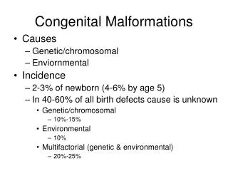
A congenital anomaly is a structural abnormality of any type that is present at birth. • Congenital anomalies may be induced by genetic or environmental factors. Most common congenital anomalies, however, show the family patterns expected of multifactorial inheritance (determined by a combination of genetic and environmental factors). • About 3% of all liveborn infants have an obvious major anomaly. • The incidence is about 6% in 2-year-olds and 8% in 5-year-olds. • Congenital anomalies may be single or multiple and of minor or major clinical significance.
During the first 2 weeks of development, teratogenic agents usually kill the embryo or have no effect. • During the organogenesis period (3rd – 8th weeks), teratogenic agents disrupt development and may cause major congenital anomalies. • During the fetal period (9th week – 9th month) teratogens may produce morphological and functional abnormalities, particularly of the brain and eyes.
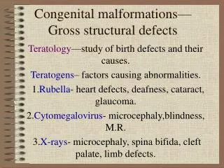
Causes of congenital anomalies 1-Genetic factors such as chromosomal abnormalities and mutant genes. 2-Environmental factors e.g.: the mother had German measles in early pregnancy will cause abnormality in the embryo. 3-Combined genetic and environmental factors (mutlifactorials factors).
Types of abnormalities 1-Malformations: this occurs during the formation of the structures of the organ (during organogenesis) results in partial or complete non formation or alterations in the normal structure. This occurs in the 3rd to the 8th week of gestation. Ex. Cleft lip and or cleft palate. 2-Disruptions: results in morphological change of the already formed structure due to exposure to destructive process. e.g.: vascular accidents leading to intestinal atresia, amniotic band disruption. 3-Deformations: due to mechanical forces that affect a part of the fetus over a long period. Ex: talipes equinovarus deformity. 4-Syndrome: is a group of anomalies occurring together due to a common cause .
The genetic factors leading to congenital anomalies may be due to chromosomal abnormalities, gene mutations or may be multifactorial. • Chromosomal abnormalities occur due to: - late maternal age at the time of pregnancy (leads to chromosomal non-disjunction), - radiation (causes chromosome deletions, translocations or breaks), - viruses as German measles, - autoimmune diseases, - and some chemical agents as anti-mitotic drugs.
- Chromosomal abnormalities are classified into numerical and structural. • Numerical chromosomal anomalies are divided into: 1- polyploidyas triploidy ( a fetus with 69 chromosomes) and tetraploidy where the fetus has 92 chromosomes. Polyploidy leads to severe congenital anomalies and early abortion.
2- Aneuploidy(one or more chromosomes is added or missed) as in: Down syndrome (trisomy 21),
Edward syndrome (trisomy 18), • Patau syndrome (trisomy 13),
Turner syndrome ((45,X or a female missing one X), and Klinefelter syndrome (47,XXY or a male person with an extra X chromosome).
Structural chromosomal anomalies include chromosomal deletion, duplication, translocation, inversion, and ring and iso chromosomes. It may also lead to severe congenital anomalies or fetal death.
Environmental factors 1) Infectious Agents: 1-Infectious agents include a number of viruses: • Rubella used to be a major problem. It causes cataract, glaucoma, heart defects and deafness. • Cytomegalovirus :The infection is often fatal and if not meningoencephalitis produce mental retardation. • Herpes simplex, varicella and human immunodeficiency viruses are other examples. 2- Toxoplasmosis 3- Syphilis : leads to congenital deafness and mental retardation.
Environmental factors Cont. 2)Radiation : Ionizing radiation kills rapidly proliferating cells, producing any type of birth defect depending upon dose and stage of development. Ex. Atomic bomb on Hiroshima and Nagasaki. Exposure of the pregnant woman to a large dose of x- ray can lead to microcephaly, spina bifida or cleft palate.
Environmental factors Cont. 3) Chemical agents: There are many dangerous drugs, if have given to the pregnant female, can produce congenital anomalies. Ex.: • Thalidomide (antinauseant sleeping pills) produce limb defects (phocomelia) and heart malformations. • Diphenylhydantoin produce facial defects and mental retardation. • Tetracycline (bone and teeth anomalies) • Aspirin may cause harm in large doses. • Cocaine cause birth defect possibly to its effect as a vasoconstrictor that cause hypoxia. • Alcohol cause fetal alcohol syndrome.
Environmental factors Cont. 5)Hormones: • Androgenic agents (synthetic progestin to prevent abortion) cause masculinization of the genitalia of female embryos. • Endocrine hormones as Diethylstilbestrol cause malformation of the uterus, uterine tubes, upper vagina, vaginal cancer and malformed testes. • Insulin which treat diabetes of the mother congenital anomalies. • Cortisone (in large doses) may cause cleft palate.
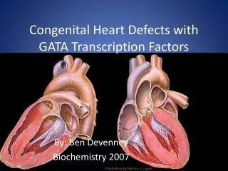
Environmental factors Cont. 6)Maternal Disease: • Diabetes cause variety of malformations as heart and neural tube defects. 7)Nutritional deficiency:particularly vitamins deficiency. 8)Heavy metals:Eg: organic mercury.
PRENATAL DIAGNOSIS • Methods of prenatal diagnosis are divided into invasive and non-invasive techniques. • Technique Time Disorders diagnosed • (in weeks) • A. Non-invasive: • Maternal serum screen: • Alpha feto protein (AFP) 16 Neural tube defects (NTD) • Triple test 16 Down syndrome • Ultrasound 18 Structural defects in many • organs as CNS, heart, • kidney, and limbs. • B. Invasive: • - Amniocentesis 14-16 Chromosomal and metabolic • abnormalities, and DNA • analysis. • - Chorionic villus sampling 10-12 As amniocentesis. • - Fetal blood sample near term As amniocentesis + blood • disorders.
U/S showing Umibilical hernia (associated with Trisomy 18 in 50% of cases)
Fetal therapy • The fetus during intrauterine life can receive treatment such as: 1- Fetal transfusion (administration of blood transfusion to the anemic fetus in thalassemia). 2- Medical treatment of thyroid dysfunction or congenital adrenal hyperplasia of the fetus. 3- Fetal surgery: is possible due to advanced ultrasound and surgical procedures eg: repair of hernia of the fetus or in case of hydrocphalus. 4- Stem cell transplantation and gene therapy: it is possible to transplant stem cells before 18 weeks of gestation of the fetus without rejection because the immunocompetence of the fetus doesn’t develop yet.