Download
1 / 22
220 likes | 284 Views
Explore the definition, etiology, and cardinal signs of inflammation, along with insights into inflammatory mediators such as cytokines, prostaglandins, leukotrienes, histamine, and platelet-activating factor presented by Prof. Dr. Baydaa H. Abdullah.

E N D
Inflammation Assist. Prof.Dr. BaydaaH.Abdullah
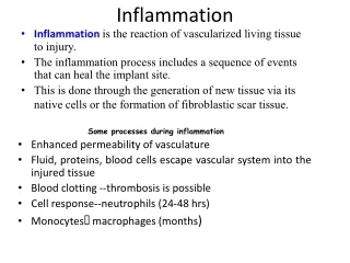
Definition • Inflammation is a non specific, localized immune reaction of the organism, which tries to localized the pathogen agent. Many consider the syndrome a self-defense mechanism. • It consist in vascular, metabolic, cellular changes, triggered by the entering of pathogen agent in healthy tissues of the body.
Etiology • The causes of inflammation are many and varied: • Exogenous causes: • Physical agents • Mechanic agents: fractures, foreign corps, sand, etc. • Thermal agents: burns, freezing • Chemical agents: toxic gases, acids, bases • Biological agents: bacteria, viruses, parasites • Endogenous causes: • Circulation disorders: thrombosis, infarction, hemorrhage • Enzymes activation – e.g. acute pancreatitis • Metabolic products deposals – uric acid, urea
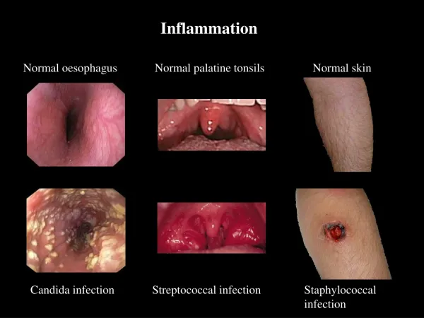
Cardinal Signs • Celsus described the local reaction of injury in terms that have come to be known as the cardinal signs of inflammation. • These signs are: • rubor (redness) • tumor (swelling) • calor (heat) • dolor (pain) • functio laesa, or loss of function (In the second century AD, the Greek physician Galen added this fifth cardinal sign)
Inflammation • The inflammatory reaction takes place at the microcirculation level and it is composed by the following changes: • Tissue damage • Cellular – vascular - cellular response • Metabolic changes • Tissue repair
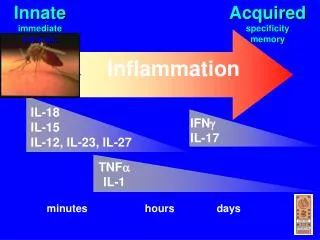
Tissue damage • Changes begin almost immediately after injury: • Because of the pathogen agent action, in the affected tissue are released mediators responsible for the following events of inflammation. • Tissue macrophages, monocytes, mast cells, platelets, and endothelial cells are able to produce a multitude of cytokines. The cytokines tissue necrosis factor-a (TNF-a) and interleukin (IL)–1 are released first and initiate several cascades.
Inflammatory Mediators • TNF-a and IL-1 are responsible for fever and the release of stress hormones (norepinephrine, vasopressin, activation of the renin-angiotensin-aldosterone system). • TNF-a and IL-1 are responsible for the synthesis of IL-6, IL-8, and interferon gamma. • Cytokines, especially IL-6, stimulate the release of acute-phase reactants such as C-reactive protein (CRP). • The proinflammatory interleukins either function directly on tissue or work via secondary mediators to activate the coagulation cascade, complement cascade, and the release of nitric oxide, platelet-activating factor, prostaglandins, and leukotrienes.
Inflammatory Mediators • Complement fragments and cytokines • It stimulates chemotaxis of neutrophils, eosinophils and monocytes; • C3a, C5a increase vascular permeability; • Cytokines • Interleukins (IL1, IL 6, IL8) • Stimulates the chemotaxis, degranulation of neutrophils and their phagocytic activity • Induce extravascularization of granulocytes • Fever • Tumor necrosis factor (TNF) and IL 8 • Leukocytosis • Fever • Stimulates prostaglandins production
Inflammatory Mediators • Prostaglandins • The prostaglandins are ubiquitous, lipid soluble molecules derived fro arachidonic acid, a fatty acid liberated from cell membrane phospholipids, through the cyclooxygenase pathway. • Prostaglandins contribute to vasodilation, capillary permeability, and the pain and fever that accompany inflammation. • The stable prostaglandins (PGE1 and PGE2) induce inflammation and potentiate the effects of histamine and other inflammatory mediators: • They cause the dilation of precapillary arterioles (edema), lower the blood pressure, modulates receptors activity and affect the phagocytic activity of leukocytes. • The prostaglandin thromboxane A2 promotes platelet aggregation and vasoconstriction.
Inflammatory Mediators • Leukotrienes • The leukotrienes are formed from arachidonic acid, but through the lipoxygenase pathway. • Histamine and leukotrienes are complementary in action in that they have similar functions. • Histamine is produced rapidly and transiently while the more potent leukotrienes are being synthesized. . • The leukotrienes also have been reported to affect the permeability of the postcapillary venules, the adhesion properties of endothelial cells, and stimulates the chemotaxis and extravascularization of neutrophils, eosinophils, and monocytes.
Inflammatory Mediators • Histamine • It is found in high concentration in platelets, basophils, and mast cells. • Causes dilation and increased permeability of capillaries (it causes dilatation of precapillary arterioles, contraction of endothelial cells and dilation of postcapillary venules). • It acts through H1 receptors.
Inflammatory Mediators • Platelet-activating factor (PAF) • It is generated from a lipid complex stored in cell membranes; • It affects a variety of cell types and induces platelet aggregation; • It activates neutrophils and is a potent eosinophil chemoattractant; • It contributes to extravascularization of plasma proteins and so, to edema.
Inflammatory Mediators • Plasma Proteases • The plasma proteases consist of: • Kinins • Bradykinin - causes increased capillary permeability (implicated in hyperthermia and redness) and pain; • Clotting factors • The clotting system contributes to the vascular phase of inflammation, mainly through fibrin peptides that are formed during the final steps of the clotting process.
The Vascular Response • Faze I = vasoconstriction (momentary constriction of small blood vessels in the area). • Vascular spasm begins very quickly (30 sec.) after the injury at it last a few minutes. • The mechanism of spasm is nervous – through catecholamine liberated from sympatic nerves endings. • Faze II = active vasodilation (through catabolism products that act through receptors and directly stimulates vascular dilation – nervous mechanism). • Dilation of arterioles and capillaries (redness = rubor); • Blood flow increases and gives pulsate sensation; • Active hyperemia in skin territory and increased metabolism leads to higher local temperature (heat = calor).
The Vascular Response • Faze III = passive vasodilation • Blood vessels in the affected area loose their reactivity to nervous and humoral stimuli and passive vasodilation occurs. • Progressively fluid move into the tissues (increased vascular permeability and structural alteration of blood vessels) and cause swelling (tumor), pain, and impaired function. • The exudation or movement of the fluid out of the capillaries and into the tissue spaces dilutes the offending agent. As fluid moves out of the capillaries, stagnation of flow and clotting of blood in the small capillaries occurs at the site of injury. • This aids in localizing the spread of infectious microorganisms, if case.
Cellular Response • The cellular response of acute inflammation is marked by movement of phagocytic white blood cells (leukocytes) into the area of injury. • Two types of leukocytes participate in the acute inflammatory response - the granulocytes and monocytes. • The sequence of events in the cellular response to inflammation includes: • pavementing • emigration • chemotaxis • phagocytosis
Pavementing • The release of chemical mediators (i.e., histamine, leukotrienes and kinins) and cytokines affects the endothelial cells of the capillaries and causes the leukocytes to increase their expression of adhesion molecules. • As this occurs, the leukocytes slow their migration and begin to marginate, or move to and along the periphery of the blood vessels.
Emigration and chemotaxis • Emigrationis a mechanism by which the leukocytes extend pseudopodia, pass through the capillary walls by ameboid movement, and migrate into the tissue spaces. • The emigration of leukocytes also may be accompanied by an escape of red blood cells. • Once they have exited the capillary, the leukocytes move through the tissue guided by secreted cytokines, bacterial and cellular debris, and complement fragments (C3a, C5a). • The process by which leukocytes migrate in response to a chemical signal is called chemotaxis.
Phagocytosis • During the next and final stage of the cellular response, the neutrophils and macrophages engulf and degrade the bacteria and cellular debris in a process called phagocytosis. • Phagocytosis involves three distinct steps: • Adherence plus opsonization • Engulfment • Intracellular killing • through enzymes, toxic oxygen and nitrogen products produced by oxygen-dependent metabolic pathways (nitric oxide, peroxyonitrites, hydrogen peroxide, and hypochlorous acid) • If the antigen is coated with antibody or complement, its adherence is increased because of binding to complement. This process of enhanced binding of an antigen caused by antibody or complement is called opsonization.
Metabolic changes • Protein metabolism • Is increased – cell destruction, metabolic products lead o increased osmotic pressure in interstitial space which attracts water and contributes to edema (swelling = tumor); • The metabolic changes, including skeletal muscle catabolism, provide amino acids that can be used in the immune response and for tissue repair; • Glucose metabolism • Anaerobe utilization of glucose is increased because of hypoxia with increased formation of lactic and pyruvic acid; • Lipid metabolism • Increased formation of ketons and fatty acids • Mineral metabolism • Increased extracellular K+ concentration • Acid – base balance • Metabolic acidosis (ketons, lactic acid)