Download
1 / 32
360 likes | 619 Views
Understand how leukocytes travel through blood vessels, inflammation processes, recirculation, and antigen collection. Learn about endothelial cells, CAMs, lymphocyte extravasation, and adhesion molecules.

E N D
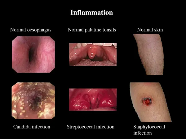
Inflammation Normal oesophagus Normal palatine tonsils Normal skin Candida infection Streptococcal infection Staphylococcal infection
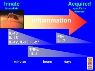
Leukocyte Migration and Inflammation • The IS relies upon the continual circulation of leukocytes through the body • For the Innate IR – a variety of lymphocytes, granulocytes, and monocytes can respond • For the Adaptive IR – lymphocytes must contact Ag in either tissue, lymph, or blood
Lymphocyte re-circulation • Lymphocytes constantly re-circulate from blood to spleen, lymph nodes, and 3° lymphoid tissues • Continual circulation provides systemic protection • A complete circuit can be performed 1-2X per day • ~1 in 105 lymphocytes can recognize a specific Ag therefore, constant circulation increases chance of lympho contacting Ag
How do leukocytes transit the bloodstream? They must bind to an endothelial cell first! • Endothelial cells exhibit ‘cell adhesion molecules’ – CAM’s • Lympho’s, granulo’s, and mono’s form receptors which bind to CAM’s
From Here to There? • Lymphoid stem cell migrate to central lymphoid organs • Mature lymphocyte migrate to peripheral lymphoid organs • Recirculation of lymphocytes • Lymphocyte migrate to the sites of inflammation
Post capillary venules in 2º lymphoid tissue HIGH ENDOTHELIAL VENULES. Specialised to allow lymphocytes and nothing else into the lymph node HEV Post capillary venules in other tissues are lined by simple squamous epithelium High endothelial venules
Recirculation Non-lymphoid cells Pass through the blood vessels in the lymph node and continue arterio-venous circulation HEV Naïve lymphoid cells Adhere to and squeeze between High Endothelial Venules (HEV), then percolate through the lymph node and exit via the efferent lymphatic vessel HEV
High endothelial venules Constitutively present in secondary lymphoid tissue Need to allow egress of naïve cells from the circulation Post-capillary venules Present in non-lymphoid tissues Role of endothelial cells in trafficking and recirculation Endothelial are involved in: Vasomotor tone, vascular permeability, regulation of coagulation, immune modulation and lymphocyte extravasation Molecules expressed by endothelial cells regulate trafficking and recirculation through lymphoid and non-lymphoid tissues
Antigen Collection • Spleen - collects antigen from the bloodstream; • Mucosa-associated lymphoid tissues (MALT) - collects antigen from the respiratory, gastrointestinal and urogenital tracts and are particularly well organized in the small intestine, in structures known as Peyer’s patches. • The lymph nodes are connected to the tissues and the bloodstream by a system of lymphatic vessels. • Afferent lymphatics drain extracellular fluid (lymph) from the tissues, including mucosal tissues, into the lymph nodes. • Efferent lymphatics carry the lymph out of the secondary lymphoid tissues and ultimately into a collecting vessel known as the thoracic duct (or for lymph nodes in the neck, the cervical duct), and thence through the heart and into the bloodstream
The four types of CAM’s • Selectins – resp. for intial contact between leukocytes and endothelial cells • Bind to specific CHO groups (i.e., mucins) • Mucins – glycosylated proteins • Bind to selectins on endothelium • Bind to other mucins (CD34 and glyCAM) on endothelium of lymph nodes
The four types of CAM’s • Integrins – heterodimer proteins formed by all leukocytes • Bind to ICAM’s along vasc. endothelium • ICAM’s – CAM’s with Ig domains on vasc. endothelia • Bind to integrins at Ig domain • MadCAM’s – have both IG and mucin-like domains; found on mucosal endothelia • Bind to integrins on lymphocytes
Selectins & addressins SELECTINS Leucocytes inc. Naive T cells:L SELECTIN Endothelial cells:P SELECTIN & E SELECTIN P selectin: Weibel-Palade bodies. E selectin: TNF & IL-1 induced A common core with different extracellular C type lectin domains that bind carbohydrates in a Ca2+ dependent manner. Each selectin binds to specific carbohydrates and is able to transduces signals into the cell VASCULAR ADDRESSINS On high endothelial venules in lymphoid tissue: Carbohydrates that “decorate” CD34 and GlyCAM-1 SialylLewisX molecules Peripheral Node addressins (PNAd) Mucosal endothelium: MAdCAM-1 Guides lymphocyte entry into lymphoid tissues
Neutrophil extravasation in inflammation Blood flow
Cell adhesions of neutrophils Rolling Adhesion Activation
Lymphocyte extravasation • Involves same 4 steps as neutrophils • Small % of endothelial cells w/i lymphoid organs exhibit “high-endothelial venules” (HEV’s) which contain many CAM’s • CAM’s function in “Homing” and “Trafficking” of lymphocytes
Associates with TcR and CD4 - phosphatase activity reduces threshold of T cell signalling CD44 CD45RA L-selectin CD45RO VLA-4 LFA-1 CD2 Naïve + + + + + - - Activated - ++ ++ ++ - + ++ Naïve cells need to access lymphoid tissue to become stimulated Memory cells need to access sites of inflammation Memory and naïve T cells
Initial contact of Naïve lymphocytes High endothelial venule cell
Trafficking, homing and adhesion Trafficking: Non-random movement of cells from tissues, blood or lymph. Includes migration to and from sites of lymphocyte maturation as well as homing. Adhesion: Binding of cells to other cells or extracellular matrix Homing: Tendency of lymphocytes activated in a particular region of the body to preferentially return to the same region Includes localisation of cells in distinct regions of lymphoid tissue.
Anti-rotavirus T cells will never be needed in the skin Anti-rotavirus T cells will be needed in the gut Anti-rotavirus T cells activated Gut Why is lymphocyte homing necessary? Tendency of lymphocytes activated in a particular region of the body to preferentially return to the same region. Gut pathogen e.g. rotavirus Response resolves, lymphocytes non-randomly redistributed
Pro-T cell migrate to thymus • Homing receptor: CD44and L-selectin expressed by pro-T cell • Adressin: EA1 molecule expressed by thymus vascular endothelial cell • And integrin α6β1、α6β4 play an important role in adhesion of pro-T cell
Lymphocyte migrate to peripheral lymphoid organs Lymphocytes migrate to lymph node • Homing receptor: L-selectin on lymphocyte • Adressin:peripheral lymphonode vascular addressin (PNAd) • LFA-1/ICAM-1、ICAM-2 and CD44/Mad molecules participate in the adhesion and penetration
Lymphocyte migrate to peripheral lymphoid organs Lymphocyte migrate to Peyer’s Patch • Homing receptor: integrin α4β7 molecule; CD44 and LFA-1 molecules • Adressin: Vascular endothelial cell of peyre’s patch specifically highly express mucosal vascular addressin (Mad); ICAM-1、ICAM-2 • Peyre’s patch means the aggregated lymphoid nodule in small intestine.
Quantitative aspects of lymphocyte migration Traffic between lymphoid/non-lymphoid tissues involves~ 5 x 10¹¹ cells per day Only ~2% (1 x 10¹⁰) of these cells are in the blood at any one time Lymphocytes only stay in the blood for ~30 minutes Circulating blood pool of lymphocytes is exchanged 48 times a day However…… Less than 10% of blood lymphocytes migrate into lymph nodes, tonsils & Peyer’s patches. ~90% of lymphocytes leave the blood to enter organs such as the liver, lung spleen and bone marrow. Traffic is 5 times faster than traffic through lymphoid tissue