Download
1 / 38
380 likes | 399 Views
Learn about the structure of nucleotides and their condensation polymers, including DNA and RNA. Understand the double helical structure of DNA and the importance of hydrogen bonding and base pairing. Discover the role of nucleic acids in carrying genetic information.

E N D
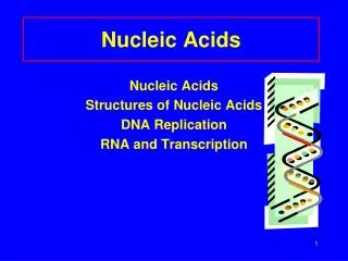
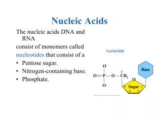
B8.1 - Describe the structure of nucleotidesand their condensation polymers(nucleic acids or polynucleotides). • Nucleic Acid – class of biopolymer, carries genetic information, also known as polynucleotides • Nucleotide – monomers of nucleic acids, combine to form polynucleotides
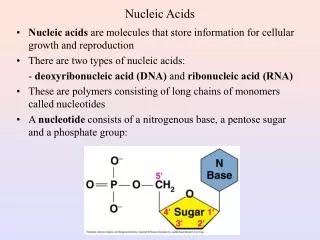
Composition of a nucleotide • Phosphate group • Pentose sugar • Organic nitrogenous base
Nucleotide Composition (con’t) • Phosphate – allows more nucleotides to be added to the chain, forming long strands; ionized, partially responsible for solubility of nucleic acids in water • Pentose Sugar – deoxyribose in DNA, ribose in RNA • Base – Continually synthesized within the cell
Bonding within nucleotides • Phosphate group is bonded covalently with the 5’ carbon of the pentose sugar • Nitrogenous base is bonded covalently with the 1’ carbon of the pentose sugar
Full nucleotide H atom H atom
Formation of polynucleotides • Condensation reaction occurs between the hydroxyl group on the 3’ carbon of one sugar and the phosphate group on the 5’ carbon of the other sugar • releases water and forms a covalent bond (known as phosphodiester bond)
Intermolecular forces • Hydrogen bonding occurs between the bases • 3 bonds occur between guanine and cytosine • 2 occur between adenine and thymine/uracil
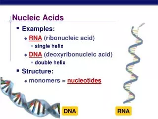
DNA RNA Deoxyribose sugar (lacks oxygen atom on C2) Adenine : Thymine bases Double helix: two polynucleotide chains held together by hydrogen bonds Stable towards enzymes and chemicals Millions of nucleotides per strand (long) Ribose (pentose) sugar Adenine : Uracil bases Typically single-stranded (but can be double in some cases) Less stable towards enzymes or chemicals 100-1,000 nucleotides per strand (short) Cytosine : Guanine bases Molecules are polynucleotides Sugar is linked to a phosphate and nitrogenous base
DNA and RNA* nucleotide structure *RNA has ribose sugar rather than deoxyribose
Nucleotide Bases • Purines are double-ringed structures • Include adenine and guanine • Pyrimidines are single-ringed • Include cytosine, thymine (in DNA), and uracil (in RNA) Purines and pyrimidines bond with one another using hydrogen bonds.
Phosphates • Link sugars together to create a strand backbone • Phosphate heads have covalent phosphodiester bonds to create a DNA or RNA strand
RNA forms • Messenger mRNA • Transfer tRNA • Ribosomal rRNA • Only RNA that can contain thymine tRNA and rRNA can be either single or double stranded. However, double-stranded RNA does not form a helix like DNA.
B.8.3 Explain the double helical structure of DNA. DNA consists of two linear polynucleotide strands which are wound together in the form of a double helix Both chains coil around the same axis Bases are on the inside of the helix Sugar-phosphate backbone on the outside Strands are anti-parallel run in opposite directions 3’ 5” and 5’ 3’
B.8.3 Explain the double helical structure of DNA. • Two chains held to together by hydrogen bonds between the bases • Double helical structure is largely due to hydrogen bonding between base pairs. • Four bases • Each have an exposed hydrogen, nitrogen, or oxygen • These can bond to other exposed hydrogen, nitrogen, or oxygen • Hydrogen bond • Special type of dipole-dipole interaction involving an attraction between an H atom bonded to an O, N, or F and an O, N, or F atom in another molecule.
Guanine Cytosine Adenine Thymine
B.8.3 Explain the double helical structure of DNA. Hydrogen bonds are weak attractions between a hydrogen atom on one side and an oxygen or nitrogen atom on the other. Hydrogen atoms of bases serve as the hydrogen bond donors The carbonyl oxygens and ring nitrogens serve as hydrogen bond acceptors The specific location of hydrogen bond donor and acceptor groups gives the bases their specificity for hydrogen bonding in unique pairs.
B.8.3 Explain the double helical structure of DNA. • Complementary base pairing • Adenine to Thymine • Cytosine to Guanine • A to T 2 hydrogen bonds • C to G 3 hydrogen bonds • One purine is paired with one pyrimidine
B.8.3 Explain the double helical structure of DNA. • Usefulness of the structure • Hydrogen bonds strongest type of the intermolecular force • Strong enough to maintain structure and keep strands together • Weak enough to separate easily • Replication can occur by breaking the hydrogen bonds
B.8.3 Explain the double helical structure of DNA. • Base stacking • Rigid bases stack on top of one another (much like stacking coins) • Purine and pyrimidines have the same width • Hydrophobic interactions and van der Waal’s forces hold the bases together
B.8.3 Explain the double helical structure of DNA. • Van der Waal’s forces draw the bases closer to each other and the DNA twists to accommodate their positions • The middle of the molecule where the bases are is hydrophobic and the polar groups are in the sugar-phosphate backbone which interacts with the aqueous solution. • The hydrophobic interactions between the bases helps to stabilize the DNA molecule.
8.4 - Describe the role of DNA as the repository of genetic information, and explain its role in protein synthesis. • DNA consists of genetic information inherited from both parents • DNA is transcribed into mRNA during transcription • mRNA is processed before leaving the nucleus • mRNA is used as a template for protein synthesis during translation
Transcription • DNA is transcribed into messenger RNA (mRNA) • RNA polymerase binds to the promoter • Unwinds the dsDNA to form an open promoter complex and initiate a transcription bubble • RNA polymerase adds nucleoside triphosphates from a 3’ to 5’ direction on the DNA (antisense) template strand • 5’ end of RNA comes out first • Nucleoside Triphosphates are being added 5’ to 3’
Transcription (con’t) • The transcription bubble moves from the DNA promoter region towards the terminator • The terminator is a sequence of nucleotides that, when transcribed, causes the RNA polymerase to detach from the DNA • The transcript carries the code of the DNA and is referred to as messenger RNA (mRNA)
Translation • Basic Information • mRNA is read in triplets by the tRNA • Triplets of mRNA are called codons • tRNA molecules have 3 bases which make anticodons • Respond to a specific amino acid that they carry • The complimentary tRNA link with mRNA and the amino acids start to line up in the right order and form peptide bonds to make a polypeptide strand
Translation • The genetic code • A triplet code • Same in all organisms - universal • Sequence of bases in DNA dictates the sequence of amino acids in all proteins via RNA • This area of biology is called the central dogma
8.5 DNA Profiling • Outline the steps involved in DNA profiling and state its use • Aim 8: include forensic and paternity tests • DNA profiling uses the techniques of genetic engineering to identify a person from a sample of their DNA • Blood, tissue, urine, bodily fluids • Used for criminal cases and paternity tests
DNA • DNA contains coding and non coding DNA • There are large portions of DNA that are identical in everyone. But some fragments of our DNA are unique to each individual • They are called the non-coding regions or “satellite DNA” • Do not code for anything and are highly repetitive in sequence (5-300 bases long) • Creates the dense and less dense regions of a DNA fingerprint used to differentiate between individuals
STRs • Thenon-coding regions that repeat are called short-tandem repeats (STRs). • Theseare looked form in multiple locations of the genome to make the tests (DNA profiling tests) more discriminating.
The Steps of DNA Profiling • Samples of cells are obtained & DNA is extracted • The sample is usually taken from blood or urine • Using restriction enzymes, the DNA is cut into small, double stranded fragments • PCR (polymerase chain reaction)is used to copy and amplify the DNA sample to produce a sufficient amount of DNA to analyze
The Steps of DNA Profiling (cont’d) 4. The fragments of DNA are then separated by gel electrophoresis into bands of different lengths • Remember: DNA fragments are negatively charged due to the phosphate groups • Place DNA in the negative side because the molecules will be attracted to the positive terminal • Shorter fragments will move further through the gel. 5. The bands are then analyzed and compared for results • The bands need to be visualized by fluorescent staining and using UV light or by using a radioactive 32P-labelledDNA probe which is exposed using X-ray film
DNA Profiling – Paternity Tests • The chromosomes of the mother and father are cut with the same restriction enzymes • A band present in the child must come from either the mother of the father • Who is the child’s father?
DNA Profiling – Paternity Tests • Example 2 : Who is Eileen’s father?
DNA Profiling – Forensic Investigations • A sample of DNA is taken from the victim or from the crime scene • DNA samples are then taken from 3 suspects • The bands of the suspects are compared to the sample at the crime scene • The victim’s DNA is also eliminated from the sample at the crime scene • Which suspect committed the crime?
sources • http://brakkeibchem1.wikispaces.com/file/view/TBD08+-+02.02.11+-+B8+Nucleic+Acids.pdf • http://www.usask.ca/education/coursework/mcvittiej/bio30unit1/overheads/1.23.htm • http://ookgm.meb.gov.tr/userfiles/file/programlar/ibo/chem_syllabusguideline(2009).pdf • http://www.microbiologyprocedure.com/genetics/chemical-nature-of-genetic-materials/molecular-structure-of-rna.htm