Download

1 / 42
480 likes | 793 Views
UJI KESEHATAN BENIH. Oleh: Irda S a fni. Outline. Pendahuluan Tujuan Uji kesehatan benih untuk jamur Deteksi bakteri patogen tular benih Deteksi virus patogen tular benih Deteksi serangga dan nematoda. Pendahuluan.

E N D
UJI KESEHATAN BENIH Oleh: Irda Safni
Outline • Pendahuluan • Tujuan • Uji kesehatan benih untuk jamur • Deteksi bakteri patogen tular benih • Deteksi virus patogen tular benih • Deteksi serangga dan nematoda
Pendahuluan • Uji kesehatan benih adalah ilmu yang menentukan ada tidaknya agen penyebab penyakit, seperti jamur, bakteri, dan virus. • Atau hama binantang seperti cacing dan serangga di dalam sampel benih.
Tujuan • Untuk menentukan status kesehatan benih yang menentukan kondisi sanitari benih untuk pasar. • Untuk mendapatkan bukti bahwa benih telah memenuhi syarat sertifikasi standar atau tidak.. • Unttuk mendapatkan bukti apakah benih telah memenuhi persyaratan karantin atau tidak.
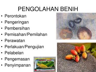
Metode uji kesehatan benih untuk jamur • Visualexamination– dengan atau tanpa stereomicroscope. • Inspeksi pada tempat yang kering bagi kehadiran kotoran seperti ergots atau skelotia lain.
Pengujian Karnalbunt (Neovassia indica) pada gandum • Benih yang terinfeksi memiliki karakteristik massa tepung hitam. • Infeksi parah – kebanyakan endosperma bersama lapisan perikarp menjadi hancur menyebabkan penampilan biji seperti kapal.
Pengujianbunt (Neovossia horrida) pada Padi • Biji dibuka dengan bantuan pisau. • Seluruh biji berubah menjadi massa hitam bertepung.
Prosedur alternatif : • Benih padidirendam di dalam larutan 0.2% sodium hidroksida selama 24 jam pada suhu 18 -25°C . • Benih yang membengkak diamati secara visual bagi warna hitam mengkilat.
Pengujian of Infeksi Loose smut pada Gandum • Benih direndam selama 24 jam pada suhu 20 °C di dalam larutan Sodium Hidroksida dan Trypan Blue. • Pisahkan embrio dengan melewati bahan yang direndam dengan air hangat (50-60°C). • Embrio dicuci di kranjang besi dan didehidrasi di dalam alkohol 95% selama 2 menit dan kemudian ditransfer ke dalam campuran laktofenol dan air.
Pemisahan lanjut embrio dapan dibuat. • Embrio ditempatkan di dalam lactophenol dan dididihkan. • Embrio diamati di mikroskop. • Jamur yang terinfeksi loose smut fungus akan tampak seperti benang hifa. • Benang coklat keemasan seperti miselium yang dipisahkan dengan ketebalan yang tidak merata dan pembengkakan diklasifikasikan sebagai embrio terinfeksi.
Pengujian UV • Toksin pada jamur memberi penampilan fluorescens. • Hal ini mewakili kehadiran jamur.
Metode BlotterMethod • Seeds are placed on moistened blotters, filter papers atleast 20mm apart. • Blotters are placed in closed containers and incubated for certain number of days and later examined for pathogens
Metode Piring Agar • Setelah benih diberi perlakuan pendahuluan (pre-treatment), benih ditempatkan pada permukaan 2% malt agar steril. • Setelah itu diinkubasi pada suhu 20-25°C selama 5-8 hari.
Metode Pencucian • Benih direndam di dalam air dengan bahan pembasah dan diguncang secara perlahan untuk melepaskan spora jamur, hifa, dst. • Sisa cairan dipindahkan dan bahan diamati.
Pengamatan Benih yang Diserap • Benih direndam di dalam air, atau cairan lain untuk membuat badan buah, gejala lebih nampak atau merangsang pembebasan spora. • Setelah penyerapan benih diamati dengan stereo mikroskop.
Metode PCR • It is a kind of molecular method. • Specific primers are used in this method. • PCR is a promising tool for distinguishing specific sequences from a complex mixture of DNA and therefore is useful for determining the presence and quantity of pathogen- specific or other unique sequences within a sample
Metode untuk mendeteksi bakteri patogen tular benih • Various methods are been developed. • Some of the methods are simple and some are specialized namely serological methods.
Growing outtest • The ‘growing out’ bioassay of a working seed sample involves the sowing of test seeds into seedlings under conditions optimal for the disease development in glass house or closed environmental chambers. • ‘Growing out’ test has been successfully used for a large number of Xanthomonads and pseudomonad’s.
Indicatortest Working seed sample is sterilized with (2.6%) sodium- hypochlorite for 15 min, and rinsed with sterile water. The seed sample is incubated for 18-24 h in sterile water. The water suspension is inoculated by infiltration into the primary leaf node of 10 day old bean seedlings. The appearance of lesions followed systemic necrosis is positive reaction.
Teknik Serologi • Serological tests are based on In vitro between antigens and antibodies. reactions • This specific recognition of antigens by antibody has offered the basic principle for the development of various serological methods for detection and identification of phytobacteria. • The washing of the working seed samples are cultured for 36 hr using sterile distilled water. • The supernatant is tested with antiserum of the suspected pathogen.
Metode Kultur • By culturing the samples on the particular media. • Presence of bacteria can be observed on the media.
Metode PCR • PCR is a promising tool for distinguishing specific sequences from a complex mixture of DNA and therefore is useful for determining the presence and quantity of pathogen- specific or other unique sequences within a sample. • It is a molecular method and it used widely used in present day.
Metode untuk mendeteksi virus patogen tular benih • BiologicalMethod • Bygrowingseeds: The seeds are examined and abnormal seeds are separated. • Both normal looking and abnormal looking seeds are grown separately and the seedings are observed for the characteristic symptom of the virus disease. • Plants are kept free of insect vectors such as mites and white fly.
DirectseedTest • Seed are examined and abnormal seeds are separated and handled separately. • Seeds are soaked in aqueous medium and then triturated. • The slurries produced are then applied to indicator or test plants. • The slurries containing virus produce symptoms typical for a given virus on the indicator plant.
Uji Serologi • Double diffusion test • Seeds to be tested are soaked in tapwater. • Seed or part (embryo) of it is triturated and triturate is transferred to a well cut in a diffusion media(agar gel). • Antiserum specific for a suspect virus in a seed is placed in separate well.
In time the virus particles(antigen) and antibody molecules diffuse towards one another. • Since diffusion is in two direction it is called Double diffusion.
Uji Radial Diffusion • Procedure consists of charging the wells with the seed or seedling preparations being tested for the presence of virus. • When virus molecules are present , they diffuse radially from the well surface they complex with antibody molecules and precipate in this region.
ELISA(Enzyme linked immunosobent assay) • In this procedure , antiserum specific for a given virus is used to coat the polystyrene plate. • Antibody molecules become absorbed. • Then seed sample is added to the plate. • It is followed by adding enzyme labelled specific antibody to the plate.
The enzyme alkaline phosphates is conjugated to the antibody molecules specific for the virus under examination. • Finally enzyme substrate is added to the plate. • Hydrolyzed substrate is determined by measuring the extinction spectrophotometrically or by visual observation.
SSEM(Serologically specific electron microscopy) • Ground seeds or homogenised preparation is applied to the electron microscope grid containing an antiserum specific for virus in question. • Samples from infected seed lots reveal the presence of virus particle characteristics for the suspect virus.
Radio Immuno Assay(RIA) • Antiserum is added to the radio labelled antigens and allowed to incubate in a centrifuge tube. • After incubation , ammonium sulphate is added for precipitation of antigen antibody complex.
After centrifugation the supernatent is discarded and the radio activity of the pallet is measured. • The pallets showing radioactivity are counted as infected. • PCR/ RT PCRis used inorder to detect the presence of Viruses
Deteksi Serangga dan Nematoda • DetectionofpresenceofInsects • Examined under magnification or stereoscopic microscope • Result is mentioned as number of insects per weight of the sample
Detectionofexternalinsectinjury • Working sample is Examined under magnification(10x) or stereoscopic microscope. • The absence of insects does not however guarantee that the seed lot is free from insect infestation, i.e internal injury to the seeds or internal infestation.
Metode Alkali atau Glyserin • Seeds are boiled in 10% NaOH solution for 10min (depending upon the kind of seeds to make them transluscent) • Alternatively the seeds can also be made transluscent by lacto- phenol solution. • Washed with water • Examined under magnification. • seeds with visible internal infestation are separated, cut opened to confirm the presence of insect or its stage and counted.
Deteksi Infestasi Nematoda • Ear cockle of wheat (Anguina tritici) • Working sample is visually examined for the presence of ear cocke galls. • (hard, small, dark purplish- black colour structures) • Galls are separated and soaked in water for 30 minutes. • and are cut in water in petridish for observing the release of nematode larvae, galls releasing nematode are counted and result is reported in percentage.
Anguinatritici Galls onwheat
Root knot Nematode of Sweet Potato (Meloidogyne incognita) • Entire submitted sample is examined for visible symptoms of nematode infestation. Galls on SweetPotato
Nematode infested tubers look ugly and disfigured. • When tubers are cut across the knot , small glistening round pin head shaped bodies are seen and such tubers are separated and the results are reported.