Download
1 / 8
80 likes | 204 Views
IMMUNOHISTOCHEMICAL EXPRESSION OF MELK IN BREAST CANCER. Manieli C*, Saccani S*, Lai ML*, Pilloni L*, Coni PP*, Senes G*, Faa G*, Van den Oord JJ°. *I Cattedra Anatomia Patologica, University of Cagliari, Italy. °University Hospitals, Katholieke Universiteit Leuven, Belgium. Background (1)

E N D
IMMUNOHISTOCHEMICAL EXPRESSION OF MELK IN BREAST CANCER Manieli C*, Saccani S*, Lai ML*, Pilloni L*, Coni PP*, Senes G*, Faa G*, Van den Oord JJ°. *I Cattedra Anatomia Patologica, University of Cagliari, Italy. °University Hospitals, Katholieke Universiteit Leuven, Belgium.
Background (1) • MELK (Maternal Embryonic Leucine Zipper Kinase) is a member of the snf1/AMPK family of serine-threonine kinases; • Its specific targets in normal cells have not been identified yet, and its role is therefore still elusive. • However, several studies reported an increased expression of MELK in various tumours compared to normal tissues.
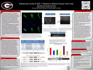
Background (2) In breast cancer cell lines and in breast tumours an elevated expression of MELK was found by semi-quantitative RT-PCR and by Western Blot analysis; MELK, by interaction with and phosphorylation of Bcl-G, a pro-apoptotic member of the Bcl-2 family, has an anti-apoptotic effect on tumour cells growth in vitro. Meng-Lay Lin et al, Breast Cancer Research 2007,9:R17.
The aim of the present study was to assess the expression of MELK by immunohistochemistry, comparing normal mammary tissue and breast tumours.
Design Immunohistochemical studies were performed on frozen sections from 17 consecutive cases of breast cancer and 4 cases of normal mammary gland, using a mouse monoclonal antibody against Melk. Immunostaining was also performed for Estrogen (ER) and Progesteron (PR) Receptors and Cerb-B2.
Results (1) • MELK was not expressed in normal mammary gland • 7 out of 17 breast tumours examined showed immunoreactivity for MELK. Normal mammary gland Invasive ductal carcinoma (IDC).
Results (2) • A cytoplasmatic staining was present in all 7 cases; 5 cases showed both cytoplasmatic and nuclear stain. • All Melk positive tumours were ER and PR positive and Cerb-B2 negative. Invasive ductal carcinoma (IDC).
Conclusions Our data show that MELK is expressed by cancer cells in a subgroup of breast tumours. In addition, MELK expression was associated with estrogen and progesteron receptor immunoreactivity. The issue of whether MELK immunostaining may have a prognostic significance in breast cancer needs further studies.