Download

1 / 72
730 likes | 779 Views
Gain insights into pediatric kidney disease patterns in Jordan, explore cultural influences, and optimize patient care strategies to avoid unnecessary investigations and save resources. Covers key topics such as congenital anomalies, antenatal diagnosis, acute kidney injury, chronic kidney disease, urinary tract infections, glomerular diseases, and more.

E N D
Approach to Pediatric Kidney Disease Kamal Akl MD Professor of Pediatrics & Pediatric Nephrology July 2019
Objectives • To familiarize medical students with the pattern of Pediatric Kidney Disease in Jordan • To approach children in a focused, geographically, and culturally oriented manner • Such approach will save time, money, and unnecessary investigations
Pediatric Kidney Disease in Jordan • Including influence of : • Cultural Habits • Religion • Economics
Approach to Pediatric Kidney Disease • Children are not small adults • Importance of the history and physical exam • Treat the Patient , not the lab • Developing Clinical sense(Sans Clinique)
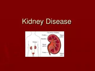
Congenital Anomalies of the Kidney and Urinary Tract(CAKUT) • Kidneys • Pelvis • Ureters • Bladder • Urethra
CAKUT • Importance of Antenatal Diagnosis
Antenatal Diagnosis • Severity of Hydronephrosis based on AP pelvic diameter: • Severe > 10 mm in 2nd trimester • Severe > 15 mm in 3rd trimester
Antenatal Diagnosis • Society for fetal urology(SFU) hydronephrosis grading system: • Grade O – no dilatation • Grade 2- Urine fills intrarenal pelvis+/- major calyces • Grade 3- all calyces are visualized • Grade 4- Grade 3+ parenchymal thininng • Nguyen HT et al • J Pediatr Urol. 2010 Jun;6(3):212-31. doi: 10.1016/j.jpurol.2010.02.205. Epub 2010 Apr 15. • The Society for Fetal Urology consensus statement on the evaluation and management of antenatal hydronephrosis.
Antenatal Diagnosis • Red flags for significant pathology: • Oligohydramnios • Additional renal or extrarenal anomalies • Sinha A • Indian J Nephrol. 2013 Mar-Apr; 23(2): 83–97.
Antenatal Diagnosis • Patients with postnatal APD exceeding 10 mm and/or SFU grade 3-4 should be screened for upper or lower urinary tract obstruction and vesicoureteric reflux (VUR). • Sinha A et al Indian J Nephrol. 2013 Mar-Apr; 23(2): 83–97.
Acute Kidney Injury • Prerenal • Intrinsic Renal • Postrenal • Causes in Jordan
Chronic Kidney Disease(CKD) • Should know • CKD stages • Updated Shwartz Formula • Causes in Jordan
CKD Stages • I • II • III • IV • V
Updated Shwartz Formula [Height(cm)/serum creatinine(mg%] x 0.413
End Stage Renal Failure(ESRF) In Jordan • Most Common causes in Jordan • CAKUT: Reflux Nephropathy • Neurogenic Bladder • Posterior Urethral Valve • Heredofamilial • Glomerular Disease • Etc • Akl KF et al • Aetiology of Paediatric End-stage Renal Failure in Jordan: A Multicentre Study. West Indian Med J. 2015 May 11;65(2):263-266.
Urinary Tract Infection(UTI) • UTI in Jordan • E coli resistance • New AAP guidelines 2011
Not all Dysuria is UTI • Causes of Dysuria: • Infectious: • APN • Cystitis • Vaginitis • Balanitis
Not all Dysuria is UTI • Noninfectious: • Dysfunctional voiding • Labial adhesions • Crystalluria Meatal stenosis Chemical irritants Trauma
Vesicoureteral Reflux • Primary • Secondary
Asymptomatic urinary abnormalities • Hematuria • Proteinuria • Hematuria+ Proteinuria
Glomerular Disease • Primary • Secondary • Jordan
Should distinguish • Between - • Glomerular vs Non Glomerular Hematuria
Should distinguish • Between • Acute Nephritis vs Nephrotic syndrome
Classification of glomerular disease • May be based on serum complement • Presentation • Histopathology
Classification based on serum complement • Hypocomplementemic : • (1) Poststrepeptococcal acute GN • (2) Mesangiocapillary GN • (3) Lupus nephritis • (4) Shunt nephritis • Normocomplementemic : Others eg MCD,FSGS , IgA N , HSP nephritis
Classification according to clinical presentation • Asymptomatic urinary abnormalities • Nephritic syndrome • Rapidly progressive glomerulonephritis • Nephrotic syndrome • Chronic glomerulonephritis • Hypertension
Classification based on histopathology • Proliferative • Nonproliferative
Primary Glomerular Disease • IgA Nephropathy • Poststreptococcal Glomerulonephritis • Jordan
Primary Glomerular Disease • Poststreptococcal AGN
Secondary Glomerular Disease • Henoch Schonlein purpura • Systemic Lupus Erythematosis • Hemolytic Uremic Syndrome
Hereditary Nephropathy • Alport syndrome • Nail Patella syndrome • New syndromes in Jordan
Acute Interstitial Nephritis • Mostly under diagnosed • Causes AKI with minimal or no proteinuria
Nephrotic Syndrome • Minimal Change Disease • Focal Segmental Glomerulosclerosis(FSGS) • Mesangioproliferative Glomerulonephritis • Mesangiocapillary Glomerulonephritis • Membranous Glomerulopathy • Seconary causes
Nephrotic syndrome • Most common causes of Steroid resistance in Jordan • FSGS • MesProliferative GN
Not all Edema is secondary to Kidney Disease • Causes of Edema: • Increased Capillary Permeability • Decreased Capillary Oncotic Pressure • Increased Plasma Volume • Lymphatic Obstruction
Should distinguish • Between • Glomerular Disease vs Tubular Disease
Tubular Disorders • Approach to Metabolic acidosis • Calculate Serum anion Gap • Urine Anion Gap
Metabolic Acidosis • Normal Anion Gap( 8-16) • Increased Anion Gap
Normal AG Metabolic Acidosis • Diarrhea • RTA
RTA • Type 1 • Type 2 • Type 4
RTA Type 1 • Ser K: low • Urine pH during acidosis :high • Ser HCO3: 10-20 • Urine pCO2: <20 • Urine citrate:low • Urolithiasis
RTA Type 2 • Ser K:Low • Urine pH during acidosis: low • Serum HCO3: 16-18 • Urine pCO2: > 20 • Urine citrate: high • Fanconi syndrome: may be present
RTA Type 4 • Ser K: High • Urine pH during acidosis: low or high • Serum HCO3: 16-22 • Urine pCO2: >20 • Urine citrate: low
Increased anion Gap Metabolic Acidosis • Poisoning: Salicylate; Toluene; ethylene Glycol;Methanol • Ketoacidosis: Diabetic; starvation • Medications • Shock • Inborn errors of Metabolism; Methanol
Metabolic Alkalosis • Chloride Responsive(U cl < 15 mEq/L) • Chloride Resistant(U cl > 20 mEq/L)
Chloride Responsive Metabolic Alkalosis • Vomiting • Cystic Fibrosis • Pyloric Stenosis