Download
1 / 22
241 likes | 653 Views
In Vivo Photoacoustic Imaging: Brain Research Application. Vassiliy Tsytsarev, E-mail: tsytsarev@umaryland.edu University of Maryland School of Medicine Most of the presented data have been obtained in: Optical Imaging Laboratory Department of Biomedical Engineering

E N D
In Vivo Photoacoustic Imaging:Brain Research Application Vassiliy Tsytsarev, E-mail: tsytsarev@umaryland.edu University of Maryland School of Medicine Most of the presented data have been obtained in: Optical Imaging Laboratory Department of Biomedical Engineering Washington University, St. Louis
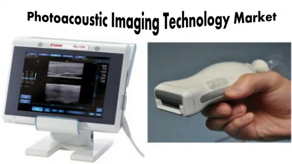
What is Photoacoustic? Photoacoustic imaging - a hybrid biomedical imaging modality, is developed based on the photoacoustic effect Photoacoustic effect – discovered by Alexander Bell in 1880; he showed that thin discs emitted sound when exposed to a beam of sunlight that was rapidly interrupted with a rotating slotted disk.
How it works? Laser Ultrasound trasducer Ultrasound
PA Imaging: three types of scanning PACT image of the cortical vasculature in a living adult intact rat Dark-field AR-PAM OR-PAM (Open Brain) (Wang et al., 2003; Stein et al., 2008)
Scheme of optical-resolution photoacoustic microscopy (OR-PAM) Wavelength-tunable laser system Condenser lens Pinhole Microscope objective Correction lens Right-angle prism Silicone oil layer Ultrasonic transducer Water tank Acoustic lens Polyethylene membrane Scanner Hu et al, 2007; (Wang et al., 2003; Stein et al., 2008)
Functional Brain Imaging: Transcranial Application In vivo PACT image of the cerebral vascular response to right-side whisker stimulation (intact rat) The hemodynamic response due to whisker stimulation is shown in blue and red and is superimposed on the cortical vascular image shown in gray. (Hu and Wang, 2009)
PA Microscopy: 2-Wavelengths Functional Imaging Hu, Maslov, Tsytsarev and Wang, 2009
Photoacoustic imaging of the vascular response Real-time monitoring of photoacoustic signals at specific excitation wavelengths reveals vascular dynamics, such as changes in blood volume and oxygen saturation, in response to electrical or physiological stimulation or epileptic seizures
Neurovascular coupling: response to electrical stimulation Electrode Neuron Astrocyte Glu GABA Glu ATP K+, NO Blood vessel Electrical stimulation may cause neurons to release various neurotransmitters (Glutamate [Glu], GABA, ATP and NO). These reactions drive the vessel to either vasoconstriction or vasodilatation
Photograph of exposed mouse brain surface with introduced microelectrode Cranial opening Microelectrode 5 mm
Photoacoustic imaging of the brain microvasculature 1.0 The oxygen saturation (SO2) mapping is shown as a superposition in color scale. A line-scan monitoring of the vascular response was performed along the dashed yellow line. SO2 0.6 1mm 400 μm Min Max Photograph of the microelectrode Optical absorption
Vascular response to electrical stimulation Transverse Axial Stimulation Axial Transverse 10 μm
50 s 50 s Vasoconstriction and vasodilatation 100 µA 150 µA 126 s 130 s Electrical stimulations 150 s 150 s 217 s 230 s 250 s 280 s 10 s 25 µm 50 µm Optical absorption Min Max
Time courses of the vessel cross-sectional area under various stimulation intensities Stimulation 2 Stimulation1 Each stimulation consisted of a train of four 0.3 ms pulses at 300 Hz Vasoconstriction and vasodilatation are observed, and the response duration is positively correlated with stimulation intensity 150 μA 300 μA 350 μA 110 μA 100 μA
Transcranial imaging of the single blood vessel (b) (a) Studied vessel medial rostral 400 μm 1.6 1.4 1.2 1.0 (c) • Transcranial images of stimulation-induced vasodilatation • Transcranial brain image • Crossectional monitoring • Time courses of the electrical-stimulation-induced vessel size 0 10 20 time (s) Stimulation
Summary • Optical-resolution photoacoustic microscopy (OR-PAM) imaged two types of vascular response to electrical stimulation: • Vasoconstriction • Vasodilatation • OR-PAM clearly and reliably imaged the vascular response to electrical stimulation at the capillary level with a temporal resolution of one second. • OR-PAM is a promising tool for in vivo studies of neurovascular coupling under a variety of experimental conditions invasively as well as transcranially
Epileptic seizures accompanied by two vessels vasodilatation (PA Imaging) EEG 10 s / 2 mV 25 μm Vessel 1 Vessel 2
Conlusions and Perspecives: • Current application: animal experiment • Clinical application: questionable • Perspective: biomarcer hybridozation